Combined Use of Residual Dipolar Couplings and Solution X-ray Scattering To Rapidly Probe Rigid-Body Conformational Transitions in a Non-phosphorylatable Active-Site Mutant of the 128 kDa Enzyme I Dimer.
Takayama, Y., Schwieters, C.D., Grishaev, A., Ghirlando, R., Clore, G.M.(2011) J Am Chem Soc 133: 424-427
- PubMed: 21162528
- DOI: https://doi.org/10.1021/ja109866w
- Primary Citation of Related Structures:
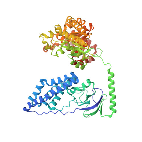
2L5H - PubMed Abstract:
The first component of the bacterial phosphotransferase system, enzyme I (EI), is a multidomain 128 kDa dimer that undergoes large rigid-body conformational transitions during the course of its catalytic cycle. Here we investigate the solution structure of a non-phosphorylatable active-site mutant in which the active-site histidine is substituted by glutamine. We show that perturbations in the relative orientations and positions of the domains and subdomains can be rapidly and reliably determined by conjoined rigid-body/torsion angle/Cartesian simulated annealing calculations driven by orientational restraints from residual dipolar couplings and shape and translation information afforded by small- and wide-angle X-ray scattering. Although histidine and glutamine are isosteric, the conformational space available to a Gln side chain is larger than that for the imidazole ring of His. An additional hydrogen bond between the side chain of Gln189 located on the EIN(α/β) subdomain and an aspartate (Asp129) on the EIN(α) subdomain results in a small (∼9°) reorientation of the EIN(α) and EIN(α/β) subdomains that is in turn propagated to a larger reorientation (∼26°) of the EIN domain relative to the EIC dimerization domain, illustrating the positional sensitivity of the EIN domain and its constituent subdomains to small structural perturbations.
Organizational Affiliation:
Laboratory of Chemical Physics, National Institute of Diabetes and Digestive and Kidney Diseases, National Institutes of Health, Bethesda, Maryland 20892-0520, United States.