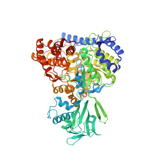
Refined solution structure of the 82-kDa enzyme malate synthase G from joint NMR and synchrotron SAXS restraints
Grishaev, A., Tugarinov, V., Kay, L.E., Trewhella, J., Bax, A.(2008) J Biomol NMR 40: 95-106
- PubMed: 18008171
- DOI: https://doi.org/10.1007/s10858-007-9211-5
- Primary Citation of Related Structures:
2JQX - PubMed Abstract:
Determination of the accurate three-dimensional structure of large proteins by NMR remains challenging due to a loss in the density of experimental restraints resulting from the often prerequisite perdeuteration. Solution small-angle scattering, which carries long-range translational information, presents an opportunity to enhance the structural accuracy of derived models when used in combination with global orientational NMR restraints such as residual dipolar couplings (RDCs) and residual chemical shift anisotropies (RCSAs). We have quantified the improvements in accuracy that can be obtained using this strategy for the 82 kDa enzyme Malate Synthase G (MSG), currently the largest single chain protein solved by solution NMR. Joint refinement against NMR and scattering data leads to an improvement in structural accuracy as evidenced by a decrease from approximately 4.5 to approximately 3.3 A of the backbone rmsd between the derived model and the high-resolution X-ray structure, PDB code 1D8C. This improvement results primarily from medium-angle scattering data, which encode the overall molecular shape, rather than the lowest angle data that principally determine the radius of gyration and the maximum particle dimension. The effect of the higher angle data, which are dominated by internal density fluctuations, while beneficial, is also found to be relatively small. Our results demonstrate that joint NMR/SAXS refinement can yield significantly improved accuracy in solution structure determination and will be especially well suited for the study of systems with limited NMR restraints such as large proteins, oligonucleotides, or their complexes.
Organizational Affiliation:
Laboratory of Chemical Physics, NIDDK, National Institutes of Health, Building 5, Bethesda, MD 20892-0520, USA. alexanderg@intra.niddk.nih.gov