


Crystal Structure of the Hypothetical Protein from Pseudomonas aeruginosa
Kim, Y., Joachimiak, A., Skarina, T., Egorova, O., Edwards, A., Savchenko, A.To be published.
Experimental Data Snapshot
wwPDB Validation 3D Report Full Report

Entity ID: 1 | |||||
|---|---|---|---|---|---|
| Molecule | Chains | Sequence Length | Organism | Details | Image |
| Hypothetical protein | 219 | Pseudomonas aeruginosa | Mutation(s): 4 | 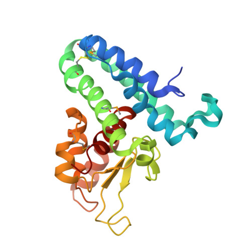 | |
UniProt | |||||
Find proteins for Q9I1P7 (Pseudomonas aeruginosa (strain ATCC 15692 / DSM 22644 / CIP 104116 / JCM 14847 / LMG 12228 / 1C / PRS 101 / PAO1)) Explore Q9I1P7 Go to UniProtKB: Q9I1P7 | |||||
Entity Groups | |||||
| Sequence Clusters | 30% Identity50% Identity70% Identity90% Identity95% Identity100% Identity | ||||
| UniProt Group | Q9I1P7 | ||||
Sequence AnnotationsExpand | |||||
| |||||
| Ligands 3 Unique | |||||
|---|---|---|---|---|---|
| ID | Chains | Name / Formula / InChI Key | 2D Diagram | 3D Interactions | |
| SO4 Query on SO4 | AA [auth H] BA [auth H] CA [auth H] I [auth A] J [auth A] | SULFATE ION O4 S QAOWNCQODCNURD-UHFFFAOYSA-L |  | ||
| GOL Query on GOL | T [auth D] | GLYCEROL C3 H8 O3 PEDCQBHIVMGVHV-UHFFFAOYSA-N |  | ||
| ACY Query on ACY | DA [auth H] L [auth A] M [auth B] P [auth C] Q [auth C] | ACETIC ACID C2 H4 O2 QTBSBXVTEAMEQO-UHFFFAOYSA-N |  | ||
| Modified Residues 1 Unique | |||||
|---|---|---|---|---|---|
| ID | Chains | Type | Formula | 2D Diagram | Parent |
| MSE Query on MSE | A, B, C, D, E A, B, C, D, E, F, G, H | L-PEPTIDE LINKING | C5 H11 N O2 Se |  | MET |
| Length ( Å ) | Angle ( ˚ ) |
|---|---|
| a = 76.424 | α = 90 |
| b = 100.458 | β = 100.33 |
| c = 140.845 | γ = 90 |
| Software Name | Purpose |
|---|---|
| REFMAC | refinement |
| SBC-Collect | data collection |
| HKL-2000 | data collection |
| HKL-2000 | data reduction |
| HKL-2000 | data scaling |
| HKL-3000 | phasing |
| SHELXCD | phasing |
| SHELXD | phasing |
| SHELXE | model building |
| MLPHARE | phasing |
| SOLVE | phasing |
| RESOLVE | phasing |
| PHENIX | phasing |
RCSB PDB (citation) is hosted by
RCSB PDB is a member of the

