Crystal Structures of the Lytic Transglycosylase MltA from N.gonorrhoeae and E.coli: Insights into Interdomain Movements and Substrate Binding.
Powell, A.J., Liu, Z.J., Nicholas, R.A., Davies, C.(2006) J Mol Biol 359: 122-136
- PubMed: 16618494
- DOI: https://doi.org/10.1016/j.jmb.2006.03.023
- Primary Citation of Related Structures:
2G5D, 2G6G, 2GAE - PubMed Abstract:
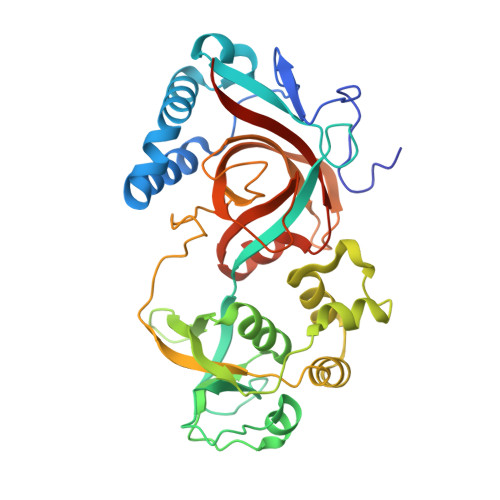
MltA is a lytic transglycosylase of Gram-negative bacteria that cleaves the beta-1,4 glycosidic linkages between N-acetylmuramic acid (MurNAc) and N-acetylglucosamine (GlcNAc) in peptidoglycan. We have determined the crystal structures of MltA from Neisseria gonorrhoeae and Escherichia coli (NgMltA and EcMltA), which have only 21.5% sequence identity. Both proteins have two main domains separated by a deep groove. Domain 1 shows structural similarity with the so-called double-psi barrel family of proteins. Comparison of the two structures reveals substantial differences in the relative positions of domains 1 and 2 such that the active site groove in NgMltA is much wider and appears more able to accommodate peptidoglycan substrate than EcMltA, suggesting that domain closure occurs after substrate binding. Docking of a peptidoglycan molecule into the structure of NgMltA reveals a number of conserved residues that are likely involved in substrate binding, including a potential binding pocket for the peptidyl moieties. This structure supports the assignment of Asp405 as the acid catalyst responsible for cleavage of the glycosidic bond. In EcMltA, the equivalent residue is Asp328, which has been identified previously. The structures also suggest a catalytic role for Asp393 (Asp317 in EcMltA) in activating the C6 hydroxyl group during formation of the 1,6-anhydro linkage. Finally, in comparison to EcMltA, NgMltA contains a unique third domain that is an insertion within domain 2. The domain is beta in structure and may mediate protein-protein interactions that are specific to peptidoglycan metabolism in N.gonorrhoeae.
Organizational Affiliation:
Department of Biochemistry & Molecular Biology, Medical University of South Carolina, Charleston, 29425, USA.