2FFU

Crystal Structure of Human ppGalNAcT-2 complexed with UDP and EA2


Experimental Data Snapshot

  • Method: X-RAY DIFFRACTION
  • Resolution: 1.64 Å
  • R-Value Free: 0.206 
  • R-Value Work: 0.178 
  • R-Value Observed: 0.179 

wwPDB Validation   3D Report Full Report


Ligand Structure Quality Assessment 


This is version 1.3 of the entry. See complete history


Literature

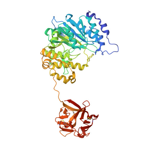
Dynamic Association between the Catalytic and Lectin Domains of Human UDP-GalNAc:Polypeptide {alpha}-N-Acetylgalactosaminyltransferase-2

Fritz, T.A.Raman, J.Tabak, L.A.

(2006) J Biol Chem 281: 8613-8619

  • DOI: https://doi.org/10.1074/jbc.M513590200
  • Primary Citation of Related Structures:  
    2FFU, 2FFV

  • PubMed Abstract: 

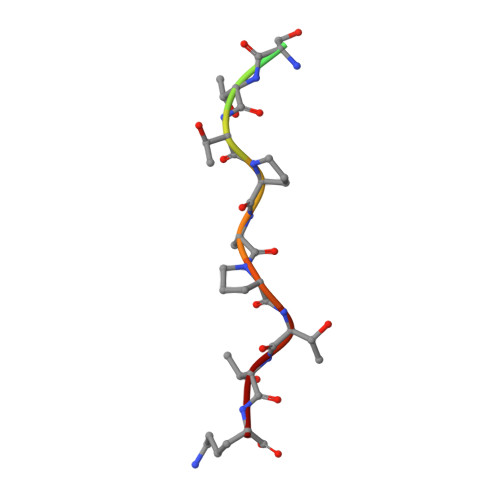
    The family of UDP-GalNAc:polypeptide alpha-N-acetylgalactosaminyltransferases (ppGalNAcTs) is unique among glycosyltransferases, containing both catalytic and lectin domains that we have previously shown to be closely associated. Here we describe the x-ray crystal structures of human ppGalNAcT-2 (hT2) bound to the product UDP at 2.75 A resolution and to UDP and an acceptor peptide substrate EA2 (PTTDSTTPAPTTK) at 1.64 A resolution. The conformations of both UDP and residues Arg362-Ser372 vary greatly between the two structures. In the hT2-UDP-EA2 complex, residues Arg362-Ser373 comprise a loop that forms a lid over UDP, sealing it in the active site, whereas in the hT2-UDP complex this loop is folded back, exposing UDP to bulk solvent. EA2 binds in a shallow groove with threonine 7 positioned consistent with in vitro data showing it to be the preferred site of glycosylation. The relative orientations of the hT2 catalytic and lectin domains differ dramatically from that of murine ppGalNAcT-1 and also vary considerably between the two hT2 complexes. Indeed, in the hT2-UDP-EA2 complex essentially no contact is made between the catalytic and lectin domains except for the peptide bridge between them. Thus, the hT2 structures reveal an unexpected flexibility between the catalytic and lectin domains and suggest a new mechanism used by hT2 to capture glycosylated substrates. Kinetic analysis of hT2 lacking the lectin domain confirmed the importance of this domain in acting on glycopeptide but not peptide substrates. The structure of the hT2-UDP-EA2 complex also resolves long standing questions regarding ppGalNAcT acceptor substrate specificity.


  • Organizational Affiliation

    Section on Biological Chemistry, NIDDK, National Institutes of Health, Bethesda, Maryland 20892, USA.


Macromolecules
Find similar proteins by:  (by identity cutoff)  |  3D Structure
Entity ID: 1
MoleculeChains Sequence LengthOrganismDetailsImage
Polypeptide N-acetylgalactosaminyltransferase 2501Homo sapiensMutation(s): 0 
Gene Names: GALNT2
EC: 2.4.1.41
UniProt & NIH Common Fund Data Resources
Find proteins for Q10471 (Homo sapiens)
Explore Q10471 
Go to UniProtKB:  Q10471
PHAROS:  Q10471
GTEx:  ENSG00000143641 
Entity Groups  
Sequence Clusters30% Identity50% Identity70% Identity90% Identity95% Identity100% Identity
UniProt GroupQ10471
Sequence Annotations
Expand
  • Reference Sequence

Find similar proteins by:  Sequence   |   3D Structure  

Entity ID: 2
MoleculeChains Sequence LengthOrganismDetailsImage
13-Peptide EA2, PTTDSTTPAPTTKB [auth P]13N/AMutation(s): 0 
UniProt
Find proteins for Q63549 (Rattus norvegicus)
Explore Q63549 
Go to UniProtKB:  Q63549
Entity Groups  
Sequence Clusters30% Identity50% Identity70% Identity90% Identity95% Identity100% Identity
UniProt GroupQ63549
Sequence Annotations
Expand
  • Reference Sequence
Small Molecules
Ligands 2 Unique
IDChains Name / Formula / InChI Key2D Diagram3D Interactions
UDP
Query on UDP

Download Ideal Coordinates CCD File 
D [auth A]URIDINE-5'-DIPHOSPHATE
C9 H14 N2 O12 P2
XCCTYIAWTASOJW-XVFCMESISA-N
MN
Query on MN

Download Ideal Coordinates CCD File 
C [auth A]MANGANESE (II) ION
Mn
WAEMQWOKJMHJLA-UHFFFAOYSA-N
Experimental Data & Validation

Experimental Data

  • Method: X-RAY DIFFRACTION
  • Resolution: 1.64 Å
  • R-Value Free: 0.206 
  • R-Value Work: 0.178 
  • R-Value Observed: 0.179 
  • Space Group: P 61
Unit Cell:
Length ( Å )Angle ( ˚ )
a = 69.344α = 90
b = 69.344β = 90
c = 169.124γ = 120
Software Package:
Software NamePurpose
CNSrefinement
HKL-2000data scaling
PHASERphasing

Structure Validation

View Full Validation Report


Ligand Structure Quality Assessment 


Entry History 

Deposition Data

  • Released Date: 2006-01-31 
  • Deposition Author(s): Fritz, T.A.

Revision History  (Full details and data files)

  • Version 1.0: 2006-01-31
    Type: Initial release
  • Version 1.1: 2008-05-01
    Changes: Version format compliance
  • Version 1.2: 2011-07-13
    Changes: Version format compliance
  • Version 1.3: 2017-10-18
    Changes: Refinement description