2F1T

Outer membrane protein OmpW


Experimental Data Snapshot

  • Method: X-RAY DIFFRACTION
  • Resolution: 3.00 Å
  • R-Value Free: 0.329 
  • R-Value Work: 0.274 
  • R-Value Observed: 0.276 

wwPDB Validation   3D Report Full Report


Ligand Structure Quality Assessment 


This is version 1.3 of the entry. See complete history


Literature

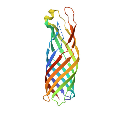
The outer membrane protein OmpW forms an eight-stranded beta-barrel with a hydrophobic channel.

Hong, H.Patel, D.R.Tamm, L.K.van den Berg, B.

(2006) J Biol Chem 281: 7568-7577

  • DOI: https://doi.org/10.1074/jbc.M512365200
  • Primary Citation of Related Structures:  
    2F1T, 2F1V

  • PubMed Abstract: 

    Escherichia coli OmpW belongs to a family of small outer membrane proteins that are widespread in Gram-negative bacteria. Their functions are unknown, but recent data suggest that they may be involved in the protection of bacteria against various forms of environmental stress. To gain insight into the function of these proteins A we have determined the crystal structure of E. coli OmpW to 2.7-A resolution. The structure shows that OmpW forms an 8-stranded beta-barrel with a long and narrow hydrophobic channel that contains a bound n-dodecyl-N,N-dimethylamine-N-oxide detergent molecule. Single channel conductance experiments show that OmpW functions as an ion channel in planar lipid bilayers. The channel activity can be blocked by the addition of n-dodecyl-N,N-dimethylamine-N-oxide. Taken together, the data suggest that members of the OmpW family could be involved in the transport of small hydrophobic molecules across the bacterial outer membrane.


  • Organizational Affiliation

    Department of Molecular Physiology and Biological Physics, University of Virginia, Charlottesville, VA 22908, USA.


Macromolecules
Find similar proteins by:  (by identity cutoff)  |  3D Structure
Entity ID: 1
MoleculeChains Sequence LengthOrganismDetailsImage
Outer membrane protein W
A, B, C
197Escherichia coli K-12Mutation(s): 0 
Gene Names: ompW
Membrane Entity: Yes 
UniProt
Find proteins for P0A915 (Escherichia coli (strain K12))
Explore P0A915 
Go to UniProtKB:  P0A915
Entity Groups  
Sequence Clusters30% Identity50% Identity70% Identity90% Identity95% Identity100% Identity
UniProt GroupP0A915
Sequence Annotations
Expand
  • Reference Sequence
Small Molecules
Ligands 3 Unique
IDChains Name / Formula / InChI Key2D Diagram3D Interactions
C8E
Query on C8E

Download Ideal Coordinates CCD File 
E [auth A]
F [auth A]
G [auth A]
H [auth A]
I [auth A]
E [auth A],
F [auth A],
G [auth A],
H [auth A],
I [auth A],
L [auth B],
M [auth B],
P [auth C]
(HYDROXYETHYLOXY)TRI(ETHYLOXY)OCTANE
C16 H34 O5
FEOZZFHAVXYAMB-UHFFFAOYSA-N
LDA
Query on LDA

Download Ideal Coordinates CCD File 
D [auth A],
K [auth B],
O [auth C]
LAURYL DIMETHYLAMINE-N-OXIDE
C14 H31 N O
SYELZBGXAIXKHU-UHFFFAOYSA-N
GOL
Query on GOL

Download Ideal Coordinates CCD File 
J [auth A],
N [auth B],
Q [auth C]
GLYCEROL
C3 H8 O3
PEDCQBHIVMGVHV-UHFFFAOYSA-N
Experimental Data & Validation

Experimental Data

  • Method: X-RAY DIFFRACTION
  • Resolution: 3.00 Å
  • R-Value Free: 0.329 
  • R-Value Work: 0.274 
  • R-Value Observed: 0.276 
  • Space Group: P 31 2 1
Unit Cell:
Length ( Å )Angle ( ˚ )
a = 82.158α = 90
b = 82.158β = 90
c = 186.234γ = 120
Software Package:
Software NamePurpose
HKL-2000data collection
SCALEPACKdata scaling
SHARPphasing
CNSrefinement
HKL-2000data reduction

Structure Validation

View Full Validation Report


Ligand Structure Quality Assessment 


Entry History 

Deposition Data

Revision History  (Full details and data files)

  • Version 1.0: 2006-01-24
    Type: Initial release
  • Version 1.1: 2008-05-01
    Changes: Version format compliance
  • Version 1.2: 2011-07-13
    Changes: Non-polymer description, Version format compliance
  • Version 1.3: 2024-02-14
    Changes: Data collection, Database references, Derived calculations