2EMT

Crystal Structure Analysis of the radixin FERM domain complexed with adhesion molecule PSGL-1


Experimental Data Snapshot

  • Method: X-RAY DIFFRACTION
  • Resolution: 2.80 Å
  • R-Value Free: 0.294 
  • R-Value Work: 0.235 
  • R-Value Observed: 0.235 

wwPDB Validation   3D Report Full Report


This is version 1.3 of the entry. See complete history


Literature

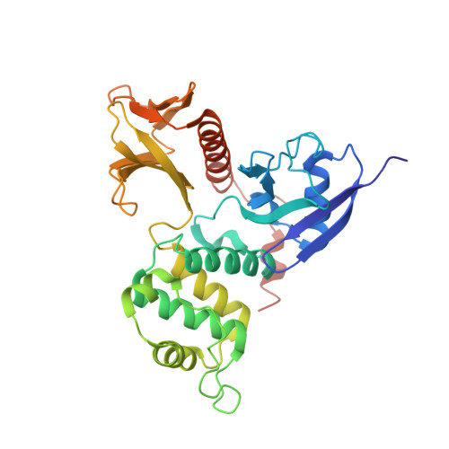
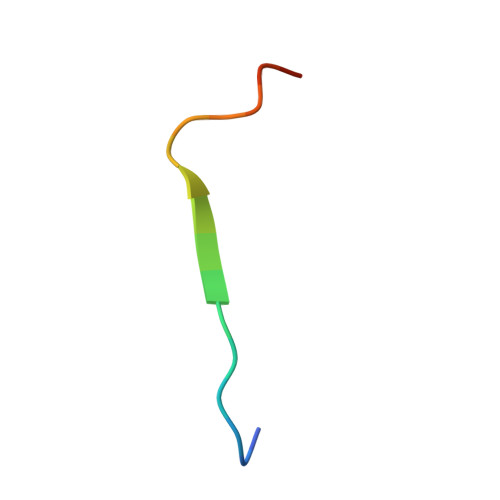
Structural basis of PSGL-1 binding to ERM proteins

Takai, Y.Kitano, K.Terawaki, S.Maesaki, R.Hakoshima, T.

(2007) Genes Cells 12: 1329-1338

  • DOI: https://doi.org/10.1111/j.1365-2443.2007.01137.x
  • Primary Citation of Related Structures:  
    2EMT

  • PubMed Abstract: 

    P-selectin glycoprotein ligand-1 (PSGL-1), an adhesion molecule with O-glycosylated extracellular sialomucins, is involved in leukocyte inflammatory responses. On activation, ezrin-radixin-moesin (ERM) proteins mediate the redistribution of PSGL-1 on polarized cell surfaces to facilitate binding to target molecules. ERM proteins recognize a short binding motif, Motif-1, conserved in cytoplasmic tails of adhesion molecules, whereas PSGL-1 lacks Motif-1 residues important for binding to ERM proteins. The crystal structure of the complex between the radixin FERM domain and a PSGL-1 juxtamembrane peptide reveals that the peptide binds the groove of FERM subdomain C by forming a beta-strand associated with strand beta5C, followed by a loop flipped out towards the solvent. The Motif-1 3(10) helix present in the FERM-ICAM-2 complex is absent in PSGL-1 given the absence of a critical Motif-1 alanine residue, and PSGL-1 reduces its contact area with subdomain C. Non-conserved positions are occupied by large residues Met9 and His8, which stabilize peptide conformation and enhance groove binding. Non-conserved residues play an important role in compensating for loss of binding energy resulting from the absence of conserved residues important for binding.


  • Organizational Affiliation

    Structural Biology Laboratory, Nara Institute of Science and Technology, Nara, Japan.


Macromolecules
Find similar proteins by:  (by identity cutoff)  |  3D Structure
Entity ID: 1
MoleculeChains Sequence LengthOrganismDetailsImage
Radixin
A, B
322Mus musculusMutation(s): 0 
UniProt & NIH Common Fund Data Resources
Find proteins for P26043 (Mus musculus)
Explore P26043 
Go to UniProtKB:  P26043
IMPC:  MGI:97887
Entity Groups  
Sequence Clusters30% Identity50% Identity70% Identity90% Identity95% Identity100% Identity
UniProt GroupP26043
Sequence Annotations
Expand
  • Reference Sequence

Find similar proteins by:  Sequence   |   3D Structure  

Entity ID: 2
MoleculeChains Sequence LengthOrganismDetailsImage
P-selectin glycoprotein ligand 1
C, D, E
18N/AMutation(s): 0 
UniProt
Find proteins for Q62170 (Mus musculus)
Explore Q62170 
Go to UniProtKB:  Q62170
Entity Groups  
Sequence Clusters30% Identity50% Identity70% Identity90% Identity95% Identity100% Identity
UniProt GroupQ62170
Sequence Annotations
Expand
  • Reference Sequence
Experimental Data & Validation

Experimental Data

  • Method: X-RAY DIFFRACTION
  • Resolution: 2.80 Å
  • R-Value Free: 0.294 
  • R-Value Work: 0.235 
  • R-Value Observed: 0.235 
  • Space Group: P 21 21 21
Unit Cell:
Length ( Å )Angle ( ˚ )
a = 80.74α = 90
b = 85.73β = 90
c = 117.751γ = 90
Software Package:
Software NamePurpose
CNSrefinement
HKL-2000data collection
HKL-2000data reduction
HKL-2000data scaling
MOLREPphasing

Structure Validation

View Full Validation Report


Entry History 

Deposition Data

Revision History  (Full details and data files)

  • Version 1.0: 2008-03-18
    Type: Initial release
  • Version 1.1: 2011-07-13
    Changes: Version format compliance
  • Version 1.2: 2017-10-11
    Changes: Refinement description
  • Version 1.3: 2023-10-25
    Changes: Data collection, Database references, Refinement description