2EGN

Crystal Structure of Tamalin PDZ Domain in Complex with mGluR5 C-terminal Peptide


Experimental Data Snapshot

  • Method: X-RAY DIFFRACTION
  • Resolution: 2.40 Å
  • R-Value Free: 0.273 
  • R-Value Work: 0.259 

wwPDB Validation   3D Report Full Report


This is version 1.4 of the entry. See complete history


Literature

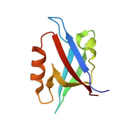
Crystal structures of autoinhibitory PDZ domain of Tamalin: implications for metabotropic glutamate receptor trafficking regulation

Sugi, T.Oyama, T.Muto, T.Nakanishi, S.Morikawa, K.Jingami, H.

(2007) EMBO J 26: 2192-2205

  • DOI: https://doi.org/10.1038/sj.emboj.7601651
  • Primary Citation of Related Structures:  
    2EGK, 2EGN, 2EGO

  • PubMed Abstract: 

    Metabotropic glutamate receptors (mGluRs) function as neuronal G-protein-coupled receptors and this requires efficient membrane targeting through associations with cytoplasmic proteins. However, the molecular mechanism regulating mGluR cell-surface trafficking remains unknown. We report here that mGluR trafficking is controlled by the autoregulatory assembly of a scaffold protein Tamalin. In the absence of mGluR, Tamalin self-assembles into autoinhibited conformations, through its PDZ domain and C-terminal intrinsic ligand motif. X-ray crystallographic analyses visualized integral parts of the oligomeric self-assemblies of Tamalin, which require not only the novel hydrophobic dimerization interface but also canonical and noncanonical PDZ/ligand autoinhibitory interactions. The mGluR cytoplasmic region can competitively bind to Tamalin at a higher concentration, disrupting weak inhibitory interactions. The atomic view of mGluR association suggests that this rearrangement is dominated by electrostatic attraction and repulsion. We also observed in mammalian cells that the association liberates the intrinsic ligand toward a motor protein receptor, thereby facilitating mGluR cell-surface trafficking. Our study suggests a novel regulatory mechanism of the PDZ domain, by which Tamalin switches between the trafficking-inhibited and -active forms depending on mGluR association.


  • Organizational Affiliation

    Department of Molecular Biology, Biomolecular Engineering Research Institute, Suita, Osaka, Japan.


Macromolecules
Find similar proteins by:  (by identity cutoff)  |  3D Structure
Entity ID: 1
MoleculeChains Sequence LengthOrganismDetailsImage
General receptor for phosphoinositides 1-associated scaffold protein96Rattus norvegicusMutation(s): 1 
UniProt
Find proteins for Q8R4T5 (Rattus norvegicus)
Explore Q8R4T5 
Go to UniProtKB:  Q8R4T5
Entity Groups  
Sequence Clusters30% Identity50% Identity70% Identity90% Identity95% Identity100% Identity
UniProt GroupQ8R4T5
Sequence Annotations
Expand
  • Reference Sequence

Find similar proteins by:  Sequence   |   3D Structure  

Entity ID: 2
MoleculeChains Sequence LengthOrganismDetailsImage
mGluR5 C-terminal peptide5Rattus norvegicusMutation(s): 0 
Sequence Annotations
Expand
  • Reference Sequence
Experimental Data & Validation

Experimental Data

  • Method: X-RAY DIFFRACTION
  • Resolution: 2.40 Å
  • R-Value Free: 0.273 
  • R-Value Work: 0.259 
  • Space Group: I 4 2 2
Unit Cell:
Length ( Å )Angle ( ˚ )
a = 75.61α = 90
b = 75.61β = 90
c = 80.185γ = 90
Software Package:
Software NamePurpose
CNSrefinement
HKL-2000data collection
HKL-2000data reduction
HKL-2000data scaling
CNSphasing

Structure Validation

View Full Validation Report


Entry History 

Deposition Data

Revision History  (Full details and data files)

  • Version 1.0: 2007-05-15
    Type: Initial release
  • Version 1.1: 2008-04-30
    Changes: Version format compliance
  • Version 1.2: 2011-07-13
    Changes: Version format compliance
  • Version 1.3: 2021-11-10
    Changes: Database references, Derived calculations
  • Version 1.4: 2023-10-25
    Changes: Data collection, Refinement description