2DQ7

Crystal Structure of Fyn kinase domain complexed with staurosporine


Experimental Data Snapshot

  • Method: X-RAY DIFFRACTION
  • Resolution: 2.80 Å
  • R-Value Free: 0.281 
  • R-Value Work: 0.255 

wwPDB Validation   3D Report Full Report


Ligand Structure Quality Assessment 


This is version 1.5 of the entry. See complete history


Literature

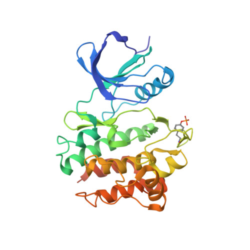
Structure of human Fyn kinase domain complexed with staurosporine.

Kinoshita, T.Matsubara, M.Ishiguro, H.Okita, K.Tada, T.

(2006) Biochem Biophys Res Commun 346: 840-844

  • DOI: https://doi.org/10.1016/j.bbrc.2006.05.212
  • Primary Citation of Related Structures:  
    2DQ7

  • PubMed Abstract: 

    The tyrosine kinase Fyn is a member of the Src kinase family. Besides the role of Fyn in T cell signal transduction in concert with Lck, its excess activity in the brain is involved with conditions such as Alzheimer's and Parkinson's diseases. Therefore, inhibition of Fyn kinase may help counteract these nervous system disorders. Here, we solved the crystal structure of the human Fyn kinase domain complexed with staurosporine, a potent kinase inhibitor, at 2.8 A resolution. Staurosporine binds to the ATP-binding site of Fyn in a similar manner as in the Lck- and Csk-complexes. The small structural differences in the staurosporine-binding and/or -unbinding region among the three kinase domains may help obtaining the selective inhibitors against the respective kinases.


  • Organizational Affiliation

    Department of Biological Science, Graduate School of Science, Osaka Prefecture University, Gakuencho 1-1, Sakai, Osaka 599-8531, Japan. kinotk@b.s.osakafu-u.ac.jp


Macromolecules
Find similar proteins by:  (by identity cutoff)  |  3D Structure
Entity ID: 1
MoleculeChains Sequence LengthOrganismDetailsImage
Proto-oncogene tyrosine-protein kinase FynA [auth X]283Homo sapiensMutation(s): 0 
EC: 2.7.10.2
UniProt & NIH Common Fund Data Resources
Find proteins for P06241 (Homo sapiens)
Explore P06241 
Go to UniProtKB:  P06241
PHAROS:  P06241
GTEx:  ENSG00000010810 
Entity Groups  
Sequence Clusters30% Identity50% Identity70% Identity90% Identity95% Identity100% Identity
UniProt GroupP06241
Sequence Annotations
Expand
  • Reference Sequence
Small Molecules
Ligands 1 Unique
IDChains Name / Formula / InChI Key2D Diagram3D Interactions
STU
Query on STU

Download Ideal Coordinates CCD File 
B [auth X]STAUROSPORINE
C28 H26 N4 O3
HKSZLNNOFSGOKW-FYTWVXJKSA-N
Modified Residues  1 Unique
IDChains TypeFormula2D DiagramParent
PTR
Query on PTR
A [auth X]L-PEPTIDE LINKINGC9 H12 N O6 PTYR
Binding Affinity Annotations 
IDSourceBinding Affinity
STU PDBBind:  2DQ7 IC50: 4.8 (nM) from 1 assay(s)
Experimental Data & Validation

Experimental Data

  • Method: X-RAY DIFFRACTION
  • Resolution: 2.80 Å
  • R-Value Free: 0.281 
  • R-Value Work: 0.255 
  • Space Group: P 32 2 1
Unit Cell:
Length ( Å )Angle ( ˚ )
a = 52.962α = 90
b = 52.962β = 90
c = 210.989γ = 120
Software Package:
Software NamePurpose
ADSCdata collection
HKL-2000data reduction
AMoREphasing
CNSrefinement
HKL-2000data scaling

Structure Validation

View Full Validation Report


Ligand Structure Quality Assessment 


Entry History 

Deposition Data

Revision History  (Full details and data files)

  • Version 1.0: 2006-07-04
    Type: Initial release
  • Version 1.1: 2008-04-30
    Changes: Version format compliance
  • Version 1.2: 2011-07-13
    Changes: Version format compliance
  • Version 1.3: 2015-04-22
    Changes: Structure summary
  • Version 1.4: 2023-10-25
    Changes: Data collection, Database references, Derived calculations, Refinement description
  • Version 1.5: 2023-11-15
    Changes: Data collection