2C62

Crystal Structure of the Human Transcription Cofactor PC4 in Complex with Single-Stranded DNA


Experimental Data Snapshot

  • Method: X-RAY DIFFRACTION
  • Resolution: 1.74 Å
  • R-Value Free: 0.240 
  • R-Value Work: 0.229 
  • R-Value Observed: 0.229 

wwPDB Validation   3D Report Full Report


This is version 1.3 of the entry. See complete history


Literature

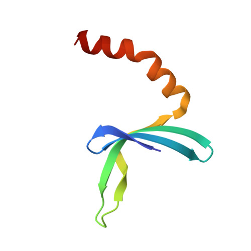
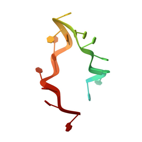
A Global Transcription Cofactor Bound to Juxtaposed Strands of Unwound DNA

Werten, S.Moras, D.

(2006) Nat Struct Mol Biol 13: 181

  • DOI: https://doi.org/10.1038/nsmb1044
  • Primary Citation of Related Structures:  
    2C62

  • PubMed Abstract: 

    The 1.74-A crystal structure of the human transcription cofactor PC4 in complex with a single-stranded 20-mer oligonucleotide reveals how symmetry-related beta-surfaces of the protein homodimer interact with juxtaposed 5-nucleotide DNA regions running in opposite directions. The structure explains high-affinity binding of PC4 to the complementary strands of unwinding duplex DNA, and it suggests the cofactor may have a role in relaxing negative supercoils or exposing unpaired bases for sequence-specific recognition by other biomolecules.


  • Organizational Affiliation

    Département de Biologie et Génomique Structurales, Institut de Génétique et de Biologie Moléculaire et Cellulaire, B.P. 10142, 67404 Illkirch Cedex, France.


Macromolecules

Find similar proteins by:  (by identity cutoff)  |  3D Structure
Entity ID: 1
MoleculeChains Sequence LengthOrganismDetailsImage
ACTIVATED RNA POLYMERASE II TRANSCRIPTIONAL COACTIVATOR P15
A, B
66Homo sapiensMutation(s): 0 
UniProt & NIH Common Fund Data Resources
Find proteins for P53999 (Homo sapiens)
Explore P53999 
Go to UniProtKB:  P53999
PHAROS:  P53999
GTEx:  ENSG00000113387 
Entity Groups  
Sequence Clusters30% Identity50% Identity70% Identity90% Identity95% Identity100% Identity
UniProt GroupP53999
Sequence Annotations
Expand
  • Reference Sequence

Find similar nucleic acids by:  Sequence   |   3D Structure  

Entity ID: 2
MoleculeChains LengthOrganismImage
5'-D(*TP*TP*TP*TP*TP*TP*TP*TP*TP*TP *TP*TP*TP*TP*TP*TP*TP*TP*TP*G)-3'20N/A
Sequence Annotations
Expand
  • Reference Sequence
Experimental Data & Validation

Experimental Data

  • Method: X-RAY DIFFRACTION
  • Resolution: 1.74 Å
  • R-Value Free: 0.240 
  • R-Value Work: 0.229 
  • R-Value Observed: 0.229 
  • Space Group: P 41 21 2
Unit Cell:
Length ( Å )Angle ( ˚ )
a = 67.242α = 90
b = 67.242β = 90
c = 131.899γ = 90
Software Package:
Software NamePurpose
CNSrefinement
DENZOdata reduction
SCALEPACKdata scaling
EPMRphasing

Structure Validation

View Full Validation Report


Entry History 

Deposition Data

Revision History  (Full details and data files)

  • Version 1.0: 2006-01-11
    Type: Initial release
  • Version 1.1: 2011-05-08
    Changes: Version format compliance
  • Version 1.2: 2011-07-13
    Changes: Version format compliance
  • Version 1.3: 2023-12-13
    Changes: Data collection, Database references, Other, Refinement description, Source and taxonomy