2B9R

Crystal Structure of Human Cyclin B1


Experimental Data Snapshot

  • Method: X-RAY DIFFRACTION
  • Resolution: 2.90 Å
  • R-Value Free: 0.308 
  • R-Value Work: 0.243 
  • R-Value Observed: 0.249 

wwPDB Validation   3D Report Full Report


This is version 1.4 of the entry. See complete history


Literature

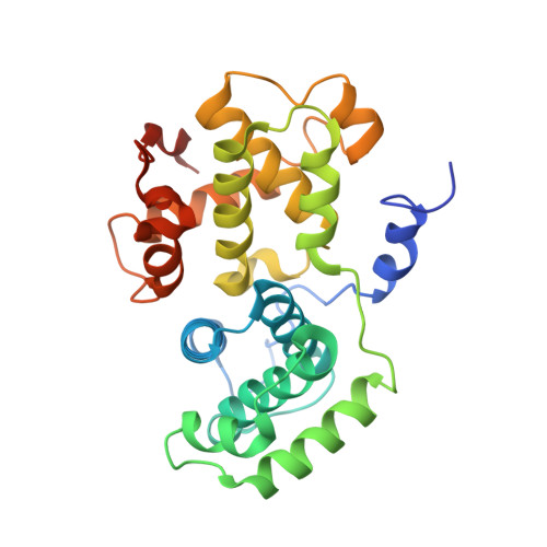
The Crystal Structure of Human Cyclin B.

Petri, E.T.Errico, A.Escobedo, L.Hunt, T.Basavappa, R.

(2007) Cell Cycle 6: 1342-1349

  • DOI: https://doi.org/10.4161/cc.6.11.4297
  • Primary Citation of Related Structures:  
    2B9R

  • PubMed Abstract: 

    Cyclin B is the key regulatory protein controlling mitosis in all eukaryotes, where it binds cyclin-dependent kinase, cdk1, forming a complex which initiates the mitotic program through phosphorylation of select proteins. Cyclin B regulates the activation, subcellular localization, and substrate specificity of cdk1, and destruction of cyclin B is necessary for mitotic exit. Overexpression of human cyclin B1 has been found in numerous cancers and has been associated with tumor aggressiveness. Here we report the crystal structure of human cyclin B1 to 2.9 A. Comparison of the structure with cyclin A and cyclin E reveals remarkably similar N-terminal cyclin box motifs but significant differences among the C-terminal cyclin box lobes. Divergence in sequence gives rise to unique interaction surfaces at the proposed cyclin B/cdk1 interface as well as the 'RxL' motif substrate binding site on cyclin B. Examination of the structure provides insight into the molecular basis for differential affinities of protein based cyclin/cdk inhibitors such as p27, substrate recognition, and cdk interaction.


  • Organizational Affiliation

    Department of Biochemistry and Biophysics, University of Rochester School of Medicine and Dentistry, Rochester, New York, USA.


Macromolecules
Find similar proteins by:  (by identity cutoff)  |  3D Structure
Entity ID: 1
MoleculeChains Sequence LengthOrganismDetailsImage
Human cyclin B1
A, B
269Homo sapiensMutation(s): 5 
UniProt & NIH Common Fund Data Resources
Find proteins for P14635 (Homo sapiens)
Explore P14635 
Go to UniProtKB:  P14635
PHAROS:  P14635
GTEx:  ENSG00000134057 
Entity Groups  
Sequence Clusters30% Identity50% Identity70% Identity90% Identity95% Identity100% Identity
UniProt GroupP14635
Sequence Annotations
Expand
  • Reference Sequence
Experimental Data & Validation

Experimental Data

  • Method: X-RAY DIFFRACTION
  • Resolution: 2.90 Å
  • R-Value Free: 0.308 
  • R-Value Work: 0.243 
  • R-Value Observed: 0.249 
  • Space Group: P 62 2 2
Unit Cell:
Length ( Å )Angle ( ˚ )
a = 117.169α = 90
b = 117.169β = 90
c = 197.685γ = 120
Software Package:
Software NamePurpose
REFMACrefinement
DENZOdata reduction
d*TREKdata scaling
PHASERphasing

Structure Validation

View Full Validation Report


Entry History 

Deposition Data

Revision History  (Full details and data files)

  • Version 1.0: 2006-10-17
    Type: Initial release
  • Version 1.1: 2008-05-01
    Changes: Version format compliance
  • Version 1.2: 2011-07-13
    Changes: Advisory, Version format compliance
  • Version 1.3: 2021-10-20
    Changes: Database references
  • Version 1.4: 2023-08-23
    Changes: Data collection, Refinement description