1WDC

SCALLOP MYOSIN REGULATORY DOMAIN


Experimental Data Snapshot

  • Method: X-RAY DIFFRACTION
  • Resolution: 2.00 Å
  • R-Value Free: 0.281 
  • R-Value Work: 0.194 
  • R-Value Observed: 0.194 

wwPDB Validation   3D Report Full Report


This is version 1.3 of the entry. See complete history


Literature

Structure of the regulatory domain of scallop myosin at 2 A resolution: implications for regulation.

Houdusse, A.Cohen, C.

(1996) Structure 4: 21-32

  • DOI: https://doi.org/10.1016/s0969-2126(96)00006-8
  • Primary Citation of Related Structures:  
    1WDC

  • PubMed Abstract: 

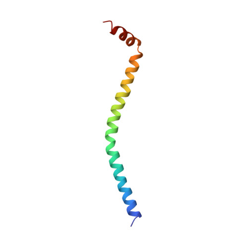
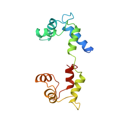
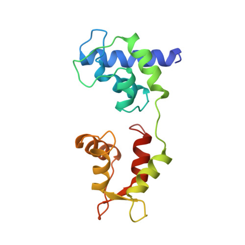
    In contrast to the myosins of vertebrate skeletal muscle, molluscan myosins are regulated molecules whose enzymatic activity is switched on by the direct binding of Ca2+. The head portion (S1) of the molecule consists of a motor domain and a regulatory domain (RD) containing a 'regulatory' and an 'essential' light chain (RLC and ELC, respectively). The structures of scallop myosin RD with bound Ca2+, as well as the S1 fragment of chicken skeletal muscle myosin, have been determined previously to 2.8 A resolution.


  • Organizational Affiliation

    Rosenstiel Basic Medical Sciences Research Center, Brandeis University, Waltham, MA 02254-9110, USA.


Macromolecules
Find similar proteins by:  (by identity cutoff)  |  3D Structure
Entity ID: 1
MoleculeChains Sequence LengthOrganismDetailsImage
SCALLOP MYOSIN64Argopecten irradiansMutation(s): 0 
UniProt
Find proteins for P24733 (Argopecten irradians)
Explore P24733 
Go to UniProtKB:  P24733
Entity Groups  
Sequence Clusters30% Identity50% Identity70% Identity90% Identity95% Identity100% Identity
UniProt GroupP24733
Sequence Annotations
Expand
  • Reference Sequence
Find similar proteins by:  (by identity cutoff)  |  3D Structure
Entity ID: 2
MoleculeChains Sequence LengthOrganismDetailsImage
SCALLOP MYOSIN156Argopecten irradiansMutation(s): 0 
UniProt
Find proteins for P13543 (Argopecten irradians)
Explore P13543 
Go to UniProtKB:  P13543
Entity Groups  
Sequence Clusters30% Identity50% Identity70% Identity90% Identity95% Identity100% Identity
UniProt GroupP13543
Sequence Annotations
Expand
  • Reference Sequence
Find similar proteins by:  (by identity cutoff)  |  3D Structure
Entity ID: 3
MoleculeChains Sequence LengthOrganismDetailsImage
SCALLOP MYOSIN156Argopecten irradiansMutation(s): 0 
UniProt
Find proteins for P07291 (Argopecten irradians)
Explore P07291 
Go to UniProtKB:  P07291
Entity Groups  
Sequence Clusters30% Identity50% Identity70% Identity90% Identity95% Identity100% Identity
UniProt GroupP07291
Sequence Annotations
Expand
  • Reference Sequence
Small Molecules
Ligands 2 Unique
IDChains Name / Formula / InChI Key2D Diagram3D Interactions
CA
Query on CA

Download Ideal Coordinates CCD File 
E [auth C]CALCIUM ION
Ca
BHPQYMZQTOCNFJ-UHFFFAOYSA-N
MG
Query on MG

Download Ideal Coordinates CCD File 
D [auth B]MAGNESIUM ION
Mg
JLVVSXFLKOJNIY-UHFFFAOYSA-N
Experimental Data & Validation

Experimental Data

  • Method: X-RAY DIFFRACTION
  • Resolution: 2.00 Å
  • R-Value Free: 0.281 
  • R-Value Work: 0.194 
  • R-Value Observed: 0.194 
  • Space Group: P 1 21 1
Unit Cell:
Length ( Å )Angle ( ˚ )
a = 52.65α = 90
b = 87.1β = 114.5
c = 55.5γ = 90
Software Package:
Software NamePurpose
X-PLORmodel building
X-PLORrefinement
R-AXISdata reduction
CCP4data reduction
X-PLORphasing

Structure Validation

View Full Validation Report


Entry History 

Deposition Data

Revision History  (Full details and data files)

  • Version 1.0: 1996-07-11
    Type: Initial release
  • Version 1.1: 2008-03-24
    Changes: Version format compliance
  • Version 1.2: 2011-07-13
    Changes: Version format compliance
  • Version 1.3: 2024-02-14
    Changes: Data collection, Database references, Derived calculations, Other