1TMH

MODULAR MUTAGENESIS OF A TIM-BARREL ENZYME: THE CRYSTAL STRUCTURE OF A CHIMERIC E. COLI TIM HAVING THE EIGHTH (BETA-ALPHA)-UNIT REPLACED BY THE EQUIVALENT UNIT OF CHICKEN TIM


Experimental Data Snapshot

  • Method: X-RAY DIFFRACTION
  • Resolution: 2.80 Å
  • R-Value Work: 0.166 
  • R-Value Observed: 0.166 

wwPDB Validation   3D Report Full Report


This is version 1.4 of the entry. See complete history


Literature

Modular mutagenesis of a TIM-barrel enzyme: the crystal structure of a chimeric E. coli TIM having the eighth beta alpha-unit replaced by the equivalent unit of chicken TIM.

Kishan, R.Zeelen, J.P.Noble, M.E.Borchert, T.V.Mainfroid, V.Goraj, K.Martial, J.A.Wierenga, R.K.

(1994) Protein Eng 7: 945-951

  • DOI: https://doi.org/10.1093/protein/7.8.945
  • Primary Citation of Related Structures:  
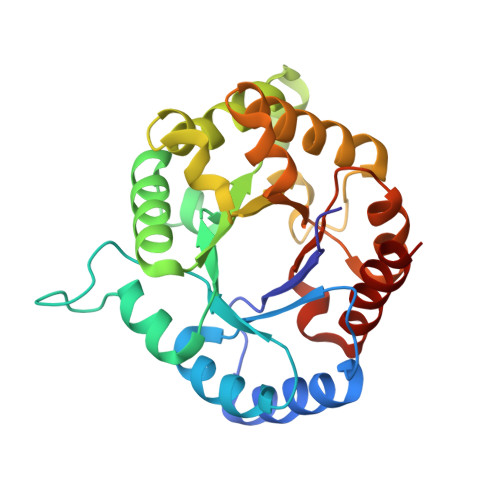
    1TMH

  • PubMed Abstract: 

    The crystal structure of a hybrid Escherichia coli triosephosphate isomerase (TIM) has been determined at 2.8 A resolution. The hybrid TIM (ETIM8CHI) was constructed by replacing the eighth beta alpha-unit of E. coli TIM with the equivalent unit of chicken TIM. This replacement involves 10 sequence changes. One of the changes concerns the mutation of a buried alanine (Ala232 in strand 8) into a phenylalanine. The ETIM8CHI structure shows that the A232F sequence change can be incorporated by a side-chain rotation of Phe224 (in helix 7). No cavities or strained dihedrals are observed in ETIM8CHI in the region near position 232, which is in agreement with the observation that ETIM8CHI and E.coli TIM have similar stabilities. The largest CA (C-alpha atom) movements, approximately 3 A, are seen for the C-terminal end of helix 8 (associated with the outward rotation of Phe224) and for the residues in the loop after helix 1 (associated with sequence changes in helix 8). From the structure it is not clear why the kcat of ETIM8CHI is 10 times lower than in wild type E.coli TIM.


  • Organizational Affiliation

    European Molecular Biology Laboratory, Heidelberg, Germany.


Macromolecules
Find similar proteins by:  (by identity cutoff)  |  3D Structure
Entity ID: 1
MoleculeChains Sequence LengthOrganismDetailsImage
TRIOSEPHOSPHATE ISOMERASE
A, B, C, D
254Escherichia coliMutation(s): 9 
EC: 5.3.1.1
UniProt
Find proteins for P00940 (Gallus gallus)
Explore P00940 
Go to UniProtKB:  P00940
Find proteins for P0A858 (Escherichia coli (strain K12))
Explore P0A858 
Go to UniProtKB:  P0A858
Entity Groups  
Sequence Clusters30% Identity50% Identity70% Identity90% Identity95% Identity100% Identity
UniProt GroupsP0A858P00940
Sequence Annotations
Expand
  • Reference Sequence
Experimental Data & Validation

Experimental Data

  • Method: X-RAY DIFFRACTION
  • Resolution: 2.80 Å
  • R-Value Work: 0.166 
  • R-Value Observed: 0.166 
  • Space Group: P 1 21 1
Unit Cell:
Length ( Å )Angle ( ˚ )
a = 63.64α = 90
b = 118.31β = 90.3
c = 58.98γ = 90
Software Package:
Software NamePurpose
X-PLORmodel building
X-PLORrefinement
X-PLORphasing

Structure Validation

View Full Validation Report


Entry History 

Deposition Data

Revision History  (Full details and data files)

  • Version 1.0: 1994-06-22
    Type: Initial release
  • Version 1.1: 2008-03-24
    Changes: Version format compliance
  • Version 1.2: 2011-07-13
    Changes: Version format compliance
  • Version 1.3: 2021-11-03
    Changes: Database references, Derived calculations, Other
  • Version 1.4: 2024-02-14
    Changes: Data collection