Following directed evolution with crystallography: structural changes observed in changing the substrate specificity of dienelactone hydrolase.
Kim, H.K., Liu, J.W., Carr, P.D., Ollis, D.L.(2005) Acta Crystallogr D Biol Crystallogr 61: 920-931
- PubMed: 15983415
- DOI: https://doi.org/10.1107/S0907444905009042
- Primary Citation of Related Structures:
1ZI6, 1ZI8, 1ZI9, 1ZIC, 1ZIX, 1ZIY, 1ZJ4, 1ZJ5 - PubMed Abstract:
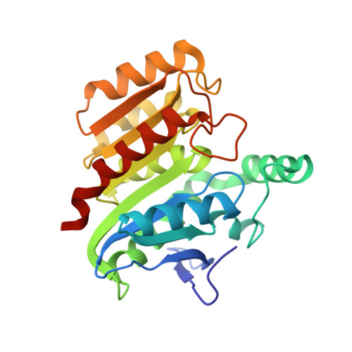
The enzyme dienelactone hydrolase (DLH) has undergone directed evolution to produce a series of mutant proteins that have enhanced activity towards the non-physiological substrates alpha-naphthyl acetate and p-nitrophenyl acetate. In terms of steady-state kinetics, the mutations caused a drop in the K(m) for the hydrolysis reaction with these two substrates. For the best mutant, there was a 5.6-fold increase in k(cat)/K(m) for the hydrolysis of alpha-naphthyl acetate and a 3.6-fold increase was observed for p-nitrophenyl acetate. For alpha-naphthyl acetate the pre-steady-state kinetics revealed that the rate constant for the formation of the covalent intermediate had increased. The mutations responsible for the rate enhancements map to the active site. The structures of the starting and mutated proteins revealed small changes in the protein owing to the mutations, while the structures of the same proteins with an inhibitor co-crystallized in the active site indicated that the mutations caused significant changes in the way the mutated proteins recognized the substrates. Within the active site of the mutant proteins, the inhibitor was rotated by about 180 degrees with respect to the orientation found in the starting enzyme. This rotation of the inhibitor caused the displacement of a large section of a loop on one side of the active site. Residues that could stabilize the transition state for the reaction were identified.
Organizational Affiliation:
Research School of Chemistry, Australian National University, Building 35, Science Road, Canberra, ACT 0200, Australia.