1YFK

Crystal structure of human B type phosphoglycerate mutase


Experimental Data Snapshot

  • Method: X-RAY DIFFRACTION
  • Resolution: 2.70 Å
  • R-Value Free: 0.255 
  • R-Value Work: 0.216 
  • R-Value Observed: 0.216 

wwPDB Validation   3D Report Full Report


This is version 1.3 of the entry. See complete history


Literature

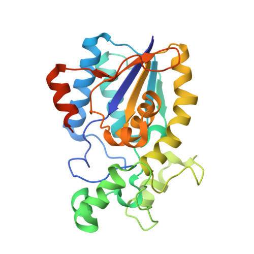
Crystal structure of human B-type phosphoglycerate mutase bound with citrate.

Wang, Y.Wei, Z.Liu, L.Cheng, Z.Lin, Y.Ji, F.Gong, W.

(2005) Biochem Biophys Res Commun 331: 1207-1215

  • DOI: https://doi.org/10.1016/j.bbrc.2005.03.243
  • Primary Citation of Related Structures:  
    1YFK, 1YJX

  • PubMed Abstract: 

    The B-type cofactor-dependent phosphoglycerate mutase (dPGM-B) catalyzes the interconversion of 2-phosphoglycerate and 3-phosphoglycerate in glycolysis and gluconeogenesis pathways using 2,3-bisphosphoglycerate as the cofactor. The crystal structures of human dPGM-B bound with citrate were determined in two crystal forms. These structures reveal a dimerization mode conserved in both of dPGM and BPGM (bisphosphoglycerate mutase), based on which a dPGM/BPGM heterodimer structure is proposed. Structural comparison supports that the conformational changes of residues 13-21 and 98-117 determine PGM/BPGM activity differences. The citrate-binding mode suggests a substrate-binding model, consistent with the structure of Escherichia coli dPGM/vanadate complex. A chloride ion was found in the center of the dimer, providing explanation for the contribution of chloride ion to dPGM activities. Based on the structural information, the possible reasons for the deficient human dPGM mutations found in some patients are also discussed.


  • Organizational Affiliation

    National Laboratory of Biomacromolecules, Institute of Biophysics, Chinese Academy of Sciences, Beijing 100101, PR China.


Macromolecules
Find similar proteins by:  (by identity cutoff)  |  3D Structure
Entity ID: 1
MoleculeChains Sequence LengthOrganismDetailsImage
Phosphoglycerate mutase 1
A, B
262Homo sapiensMutation(s): 0 
EC: 5.4.2.1 (PDB Primary Data), 5.4.2.4 (PDB Primary Data), 3.1.3.13 (PDB Primary Data)
UniProt & NIH Common Fund Data Resources
Find proteins for P18669 (Homo sapiens)
Explore P18669 
Go to UniProtKB:  P18669
PHAROS:  P18669
GTEx:  ENSG00000171314 
Entity Groups  
Sequence Clusters30% Identity50% Identity70% Identity90% Identity95% Identity100% Identity
UniProt GroupP18669
Sequence Annotations
Expand
  • Reference Sequence
Experimental Data & Validation

Experimental Data

  • Method: X-RAY DIFFRACTION
  • Resolution: 2.70 Å
  • R-Value Free: 0.255 
  • R-Value Work: 0.216 
  • R-Value Observed: 0.216 
  • Space Group: P 1 21 1
Unit Cell:
Length ( Å )Angle ( ˚ )
a = 42.861α = 90
b = 65.745β = 94.36
c = 124.812γ = 90
Software Package:
Software NamePurpose
CNSrefinement
DENZOdata reduction
SCALEPACKdata scaling
AMoREphasing

Structure Validation

View Full Validation Report


Entry History 

Deposition Data

Revision History  (Full details and data files)

  • Version 1.0: 2005-05-17
    Type: Initial release
  • Version 1.1: 2008-04-30
    Changes: Version format compliance
  • Version 1.2: 2011-07-13
    Changes: Version format compliance
  • Version 1.3: 2023-10-25
    Changes: Data collection, Database references, Derived calculations, Refinement description