1WQV

Human Factor Viia-Tissue Factor Complexed with propylsulfonamide-D-Thr-Met-p-aminobenzamidine


Experimental Data Snapshot

  • Method: X-RAY DIFFRACTION
  • Resolution: 2.50 Å
  • R-Value Free: 0.272 
  • R-Value Work: 0.218 

wwPDB Validation   3D Report Full Report


Ligand Structure Quality Assessment 


This is version 1.5 of the entry. See complete history


Literature

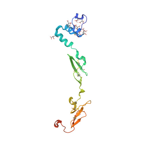
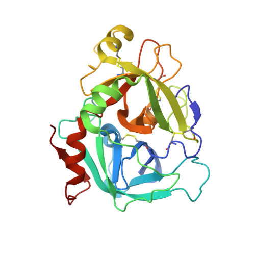
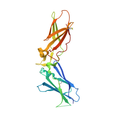
Crystal structure of human factor VIIa/tissue factor in complex with peptide mimetic inhibitor

Kadono, S.Sakamoto, A.Kikuchi, Y.Oh-eda, M.Yabuta, N.Koga, T.Hattori, K.Shiraishi, T.Haramura, M.Kodama, H.Esaki, T.Sato, H.Watanabe, Y.Itoh, S.Ohta, M.Kozono, T.

(2004) Biochem Biophys Res Commun 324: 1227-1233

  • DOI: https://doi.org/10.1016/j.bbrc.2004.09.182
  • Primary Citation of Related Structures:  
    1WQV

  • PubMed Abstract: 

    The 3D structure of human factor VIIa/soluble tissue factor in complex with a peptide mimetic inhibitor, propylsulfonamide-D-Thr-Met-p-aminobenzamidine, is determined by X-ray crystallography. As compared with the interactions between thrombin and thrombin inhibitors, the interactions at S2 and S3 sites characteristic of factor VIIa and factor VIIa inhibitors are revealed. The S2 site has a small pocket, which is filled by the hydrophobic methionine side chain in P2. The small S3 site fits the small size residue, D-threonine in P3. The structural data and SAR data of the peptide mimetic inhibitor show that these interactions in the S2 and S3 sites play an important role for the improvement of selectivity versus thrombin. The results will provide valuable information for the structure-based drug design of specific inhibitors for FVIIa/TF.


  • Organizational Affiliation

    Fuji Gotemba Research Labs, Chugai Pharmaceutical Co., Ltd., 1-135 Komakado, Gotemba, Shizuoka 412-8513, Japan. kadonosuj@chugai-pharm.co.jp


Macromolecules
Find similar proteins by:  (by identity cutoff)  |  3D Structure
Entity ID: 1
MoleculeChains Sequence LengthOrganismDetailsImage
Coagulation factor VIIA [auth L]152Homo sapiensMutation(s): 0 
EC: 3.4.21.21
UniProt & NIH Common Fund Data Resources
Find proteins for P08709 (Homo sapiens)
Explore P08709 
Go to UniProtKB:  P08709
PHAROS:  P08709
GTEx:  ENSG00000057593 
Entity Groups  
Sequence Clusters30% Identity50% Identity70% Identity90% Identity95% Identity100% Identity
UniProt GroupP08709
Sequence Annotations
Expand
  • Reference Sequence
Find similar proteins by:  (by identity cutoff)  |  3D Structure
Entity ID: 2
MoleculeChains Sequence LengthOrganismDetailsImage
Coagulation factor VIIB [auth H]254Homo sapiensMutation(s): 0 
EC: 3.4.21.21
UniProt & NIH Common Fund Data Resources
Find proteins for P08709 (Homo sapiens)
Explore P08709 
Go to UniProtKB:  P08709
PHAROS:  P08709
GTEx:  ENSG00000057593 
Entity Groups  
Sequence Clusters30% Identity50% Identity70% Identity90% Identity95% Identity100% Identity
UniProt GroupP08709
Sequence Annotations
Expand
  • Reference Sequence
Find similar proteins by:  (by identity cutoff)  |  3D Structure
Entity ID: 3
MoleculeChains Sequence LengthOrganismDetailsImage
Tissue factorC [auth T]218Homo sapiensMutation(s): 0 
UniProt & NIH Common Fund Data Resources
Find proteins for P13726 (Homo sapiens)
Explore P13726 
Go to UniProtKB:  P13726
PHAROS:  P13726
GTEx:  ENSG00000117525 
Entity Groups  
Sequence Clusters30% Identity50% Identity70% Identity90% Identity95% Identity100% Identity
UniProt GroupP13726
Sequence Annotations
Expand
  • Reference Sequence
Small Molecules
Ligands 4 Unique
IDChains Name / Formula / InChI Key2D Diagram3D Interactions
PSM
Query on PSM

Download Ideal Coordinates CCD File 
O [auth H]N-[DIHYDROXY(PROPYL)-LAMBDA~4~-SULFANYL]THREONYL-N~1~-{4-[AMINO(IMINO)METHYL]BENZYL}METHIONINAMIDE
C20 H33 N5 O5 S2
AIIJKVORRBMJHS-RRQGHBQHSA-N
BGC
Query on BGC

Download Ideal Coordinates CCD File 
D [auth L]beta-D-glucopyranose
C6 H12 O6
WQZGKKKJIJFFOK-VFUOTHLCSA-N
FUC
Query on FUC

Download Ideal Coordinates CCD File 
E [auth L]alpha-L-fucopyranose
C6 H12 O5
SHZGCJCMOBCMKK-SXUWKVJYSA-N
CA
Query on CA

Download Ideal Coordinates CCD File 
F [auth L]
G [auth L]
H [auth L]
I [auth L]
J [auth L]
F [auth L],
G [auth L],
H [auth L],
I [auth L],
J [auth L],
K [auth L],
L,
M [auth L],
N [auth H]
CALCIUM ION
Ca
BHPQYMZQTOCNFJ-UHFFFAOYSA-N
Modified Residues  1 Unique
IDChains TypeFormula2D DiagramParent
CGU
Query on CGU
A [auth L]L-PEPTIDE LINKINGC6 H9 N O6GLU
Binding Affinity Annotations 
IDSourceBinding Affinity
PSM PDBBind:  1WQV IC50: 130 (nM) from 1 assay(s)
Binding MOAD:  1WQV IC50: 130 (nM) from 1 assay(s)
Experimental Data & Validation

Experimental Data

  • Method: X-RAY DIFFRACTION
  • Resolution: 2.50 Å
  • R-Value Free: 0.272 
  • R-Value Work: 0.218 
  • Space Group: P 21 21 21
Unit Cell:
Length ( Å )Angle ( ˚ )
a = 71.39α = 90
b = 82.42β = 90
c = 123.51γ = 90
Software Package:
Software NamePurpose
CNXrefinement
DENZOdata reduction
SCALEPACKdata scaling
CNXphasing

Structure Validation

View Full Validation Report


Ligand Structure Quality Assessment 


Entry History 

Deposition Data

Revision History  (Full details and data files)

  • Version 1.0: 2005-10-02
    Type: Initial release
  • Version 1.1: 2008-04-30
    Changes: Version format compliance
  • Version 1.2: 2011-07-13
    Changes: Atomic model, Database references, Derived calculations, Non-polymer description, Structure summary, Version format compliance
  • Version 1.3: 2020-07-29
    Type: Remediation
    Reason: Carbohydrate remediation
    Changes: Data collection, Derived calculations, Structure summary
  • Version 1.4: 2023-10-25
    Changes: Data collection, Database references, Refinement description, Structure summary
  • Version 1.5: 2023-11-15
    Changes: Data collection