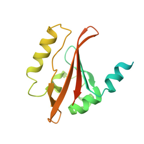
1V9Z

Crystal Structure of the heme PAS sensor domain of Ec DOS (Ferrous Form)


Experimental Data Snapshot

  • Method: X-RAY DIFFRACTION
  • Resolution: 1.90 Å
  • R-Value Free: 0.287 
  • R-Value Work: 0.244 

wwPDB Validation   3D Report Full Report


Ligand Structure Quality Assessment 


This is version 1.3 of the entry. See complete history


Literature

A redox-controlled molecular switch revealed by the crystal structure of a bacterial heme PAS sensor.

Kurokawa, H.Lee, D.S.Watanabe, M.Sagami, I.Mikami, B.Raman, C.S.Shimizu, T.

(2004) J Biol Chem 279: 20186-20193

  • DOI: https://doi.org/10.1074/jbc.M314199200
  • Primary Citation of Related Structures:  
    1V9Y, 1V9Z

  • PubMed Abstract: 

    PAS domains, which have been identified in over 1100 proteins from all three kingdoms of life, convert various input stimuli into signals that propagate to downstream components by modifying protein-protein interactions. One such protein is the Escherichia coli redox sensor, Ec DOS, a phosphodiesterase that degrades cyclic adenosine monophosphate in a redox-dependent manner. Here we report the crystal structures of the heme PAS domain of Ec DOS in both inactive Fe(3+) and active Fe(2+) forms at 1.32 and 1.9 A resolution, respectively. The protein folds into a characteristic PAS domain structure and forms a homodimer. In the Fe(3+) form, the heme iron is ligated to a His-77 side chain and a water molecule. Heme iron reduction is accompanied by heme-ligand switching from the water molecule to a side chain of Met-95 from the FG loop. Concomitantly, the flexible FG loop is significantly rigidified, along with a change in the hydrogen bonding pattern and rotation of subunits relative to each other. The present data led us to propose a novel redox-regulated molecular switch in which local heme-ligand switching may trigger a global "scissor-type" subunit movement that facilitates catalytic control.


  • Organizational Affiliation

    Institute of Multidisciplinary Research for Advanced Materials, Tohoku University, 2-1-1 Katahira, Aoba-ku, Sendai, Miyagi 980-8577, Japan. kurokawa@tagen.tohoku.ac.jp


Macromolecules
Find similar proteins by:  (by identity cutoff)  |  3D Structure
Entity ID: 1
MoleculeChains Sequence LengthOrganismDetailsImage
Heme pas sensor protein
A, B
167Escherichia coli K-12Mutation(s): 0 
Gene Names: DOS
UniProt
Find proteins for P76129 (Escherichia coli (strain K12))
Explore P76129 
Go to UniProtKB:  P76129
Entity Groups  
Sequence Clusters30% Identity50% Identity70% Identity90% Identity95% Identity100% Identity
UniProt GroupP76129
Sequence Annotations
Expand
  • Reference Sequence
Small Molecules
Ligands 1 Unique
IDChains Name / Formula / InChI Key2D Diagram3D Interactions
HEM
Query on HEM

Download Ideal Coordinates CCD File 
C [auth A],
D [auth B]
PROTOPORPHYRIN IX CONTAINING FE
C34 H32 Fe N4 O4
KABFMIBPWCXCRK-RGGAHWMASA-L
Experimental Data & Validation

Experimental Data

Unit Cell:
Length ( Å )Angle ( ˚ )
a = 46.4α = 90
b = 69.18β = 90
c = 82.45γ = 90
Software Package:
Software NamePurpose
CNSrefinement
MOSFLMdata reduction
CCP4data scaling
CNSphasing

Structure Validation

View Full Validation Report


Ligand Structure Quality Assessment 


Entry History 

Deposition Data

Revision History  (Full details and data files)

  • Version 1.0: 2004-05-25
    Type: Initial release
  • Version 1.1: 2008-04-27
    Changes: Version format compliance
  • Version 1.2: 2011-07-13
    Changes: Source and taxonomy, Version format compliance
  • Version 1.3: 2023-12-27
    Changes: Data collection, Database references, Derived calculations