1U6H

Vinculin head (0-258) in complex with the talin vinculin binding site 2 (849-879)


Experimental Data Snapshot

  • Method: X-RAY DIFFRACTION
  • Resolution: 2.38 Å
  • R-Value Free: 0.322 
  • R-Value Work: 0.242 
  • R-Value Observed: 0.246 

wwPDB Validation   3D Report Full Report


This is version 1.4 of the entry. See complete history


Literature

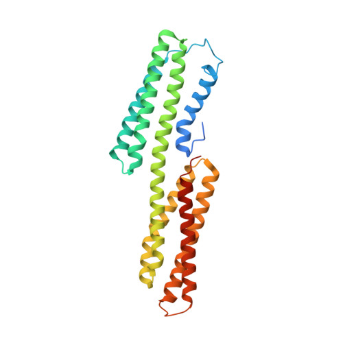
A vinculin binding domain from the talin rod unfolds to form a complex with the vinculin head.

Fillingham, I.Gingras, A.R.Papagrigoriou, E.Patel, B.Emsley, J.Critchley, D.R.Roberts, G.C.Barsukov, I.L.

(2005) Structure 13: 65-74

  • DOI: https://doi.org/10.1016/j.str.2004.11.006
  • Primary Citation of Related Structures:  
    1U6H, 1U89

  • PubMed Abstract: 

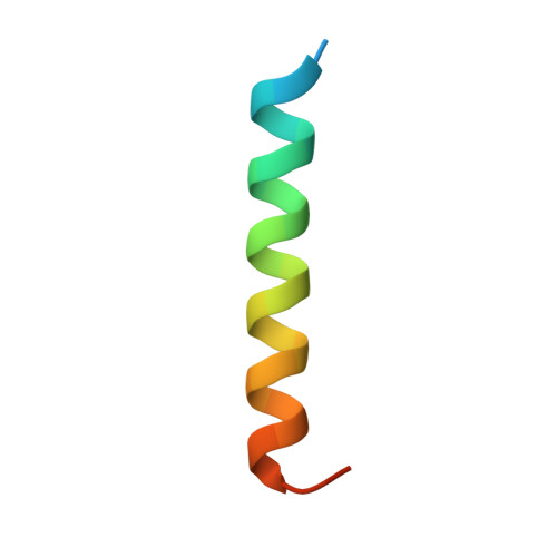
    The cytoskeletal protein talin plays a key role in activating integrins and in coupling them to the actin cytoskeleton. Its N-terminal globular head, which binds beta integrins, is linked to an extended rod having a C-terminal actin binding site and several vinculin binding sites (VBSs). The NMR structure of residues 755-889 of the rod (containing a VBS) is shown to be an amphipathic four-helix bundle with a left-handed topology. A talin peptide corresponding to the VBS binds the vinculin head; the X-ray crystallographic structure of this complex shows that the residues which interact with vinculin are buried in the hydrophobic core of the talin fragment. NMR shows that the interaction involves a major structural change in the talin fragment, including unfolding of one of its helices, making the VBS accessible to vinculin. Interestingly, the talin 755-889 fragment binds more than one vinculin head molecule, suggesting that the talin rod may contain additional as yet unrecognized VBSs.


  • Organizational Affiliation

    Department of Biochemistry, University of Leicester, Leicester LE1 7RH, UK.


Macromolecules
Find similar proteins by:  (by identity cutoff)  |  3D Structure
Entity ID: 1
MoleculeChains Sequence LengthOrganismDetailsImage
Vinculin280Gallus gallusMutation(s): 0 
Gene Names: VCLVINC1
UniProt
Find proteins for P12003 (Gallus gallus)
Explore P12003 
Go to UniProtKB:  P12003
Entity Groups  
Sequence Clusters30% Identity50% Identity70% Identity90% Identity95% Identity100% Identity
UniProt GroupP12003
Sequence Annotations
Expand
  • Reference Sequence
Find similar proteins by:  (by identity cutoff)  |  3D Structure
Entity ID: 2
MoleculeChains Sequence LengthOrganismDetailsImage
Talin31N/AMutation(s): 0 
UniProt
Find proteins for P54939 (Gallus gallus)
Explore P54939 
Go to UniProtKB:  P54939
Entity Groups  
Sequence Clusters30% Identity50% Identity70% Identity90% Identity95% Identity100% Identity
UniProt GroupP54939
Sequence Annotations
Expand
  • Reference Sequence
Experimental Data & Validation

Experimental Data

  • Method: X-RAY DIFFRACTION
  • Resolution: 2.38 Å
  • R-Value Free: 0.322 
  • R-Value Work: 0.242 
  • R-Value Observed: 0.246 
  • Space Group: P 21 21 2
Unit Cell:
Length ( Å )Angle ( ˚ )
a = 52.122α = 90
b = 69.154β = 90
c = 96.162γ = 90
Software Package:
Software NamePurpose
REFMACrefinement
DENZOdata reduction
SCALEPACKdata scaling
AMoREphasing

Structure Validation

View Full Validation Report


Entry History 

Deposition Data

Revision History  (Full details and data files)

  • Version 1.0: 2005-01-18
    Type: Initial release
  • Version 1.1: 2008-04-30
    Changes: Version format compliance
  • Version 1.2: 2011-07-13
    Changes: Version format compliance
  • Version 1.3: 2024-02-14
    Changes: Data collection, Database references
  • Version 1.4: 2024-04-03
    Changes: Refinement description