1TD9

Crystal Structure of a Phosphotransacetylase from Bacillus subtilis


Experimental Data Snapshot

  • Method: X-RAY DIFFRACTION
  • Resolution: 2.75 Å
  • R-Value Free: 0.297 
  • R-Value Work: 0.268 
  • R-Value Observed: 0.268 

wwPDB Validation   3D Report Full Report


This is version 1.4 of the entry. See complete history


Literature

Crystal structures of a phosphotransacetylase from Bacillus subtilis and its complex with acetyl phosphate

Xu, Q.S.Jancarik, J.Lou, Y.Kuznetsova, K.Yakunin, A.F.Yokota, H.Adams, P.Kim, R.Kim, S.-H.

(2005) J Struct Funct Genomics 6: 269-279

  • DOI: https://doi.org/10.1007/s10969-005-9001-9
  • Primary Citation of Related Structures:  
    1TD9, 1XCO

  • PubMed Abstract: 

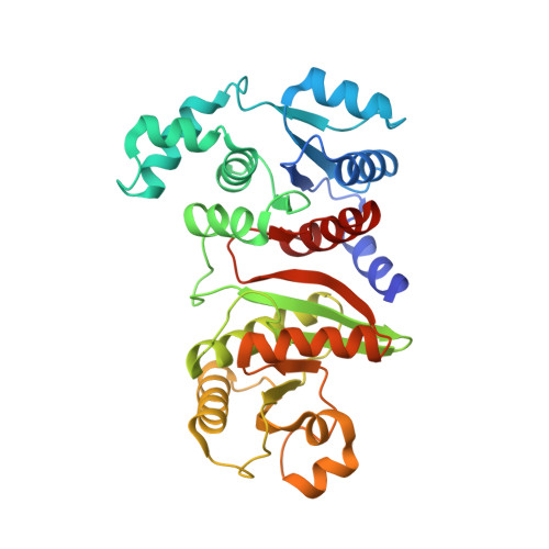
    Phosphotransacetylase (Pta) [EC 2.3.1.8] plays a major role in acetate metabolism by catalyzing the reversible transfer of the acetyl group between coenzyme A (CoA) and orthophosphate: CH(3)COSCoA+HPO(4)(2-)<-->CH(3)COOPO(3)(2-) +CoASH. In this study, we report the crystal structures of Pta from Bacillus subtilis at 2.75 A resolution and its complex with acetyl phosphate, one of its substrates, at 2.85 A resolution. In addition, the Pta activity of the enzyme has been assayed. The enzyme folds into an alpha/beta architecture with two domains separated by a prominent cleft, very similar to two other known Pta structures. The enzyme-acetyl phosphate complex structure reveals a few potential substrate binding sites. Two of them are located in the middle of the interdomain cleft: each one is surrounded by a region of strictly and highly conserved residues. High structural similarities are found with 4-hydroxythreonine-4-phosphate dehydrogenase (PdxA), and isocitrate and isopropylmalate dehydrogenases, all of which utilize NADP+ as their cofactor, which binds in the interdomain cleft. Their substrate binding sites are close to the acetyl phosphate binding sites of Pta in the cleft as well. These results suggest that the CoA is likely to bind to the interdomain cleft of Pta in a similar way as NADP+ binds to the other three enzymes.


  • Organizational Affiliation

    Berkeley Structural Genomics Center, Physical Biosciences Division, Lawrence Berkeley National Laboratory, Berkeley, CA 94720, USA.


Macromolecules
Find similar proteins by:  (by identity cutoff)  |  3D Structure
Entity ID: 1
MoleculeChains Sequence LengthOrganismDetailsImage
Phosphate acetyltransferase
A, B, C, D, E
A, B, C, D, E, F
329Bacillus subtilisMutation(s): 0 
Gene Names: PTAIPA-88DBSU37660
EC: 2.3.1.8
UniProt
Find proteins for P39646 (Bacillus subtilis (strain 168))
Explore P39646 
Go to UniProtKB:  P39646
Entity Groups  
Sequence Clusters30% Identity50% Identity70% Identity90% Identity95% Identity100% Identity
UniProt GroupP39646
Sequence Annotations
Expand
  • Reference Sequence
Small Molecules
Ligands 1 Unique
IDChains Name / Formula / InChI Key2D Diagram3D Interactions
SO4
Query on SO4

Download Ideal Coordinates CCD File 
AA [auth E]
BA [auth E]
CA [auth F]
DA [auth F]
G [auth A]
AA [auth E],
BA [auth E],
CA [auth F],
DA [auth F],
G [auth A],
H [auth A],
I [auth A],
J [auth A],
K [auth A],
L [auth B],
M [auth B],
N [auth B],
O [auth B],
P [auth C],
Q [auth C],
R [auth C],
S [auth C],
T [auth C],
U [auth D],
V [auth D],
W [auth D],
X [auth D],
Y [auth E],
Z [auth E]
SULFATE ION
O4 S
QAOWNCQODCNURD-UHFFFAOYSA-L
Experimental Data & Validation

Experimental Data

  • Method: X-RAY DIFFRACTION
  • Resolution: 2.75 Å
  • R-Value Free: 0.297 
  • R-Value Work: 0.268 
  • R-Value Observed: 0.268 
  • Space Group: P 64 2 2
Unit Cell:
Length ( Å )Angle ( ˚ )
a = 185.207α = 90
b = 185.207β = 90
c = 259.414γ = 120
Software Package:
Software NamePurpose
CNSrefinement
HKL-2000data reduction
SCALEPACKdata scaling
CNSphasing

Structure Validation

View Full Validation Report


Entry History 

Deposition Data

Revision History  (Full details and data files)

  • Version 1.0: 2004-12-07
    Type: Initial release
  • Version 1.1: 2008-04-30
    Changes: Version format compliance
  • Version 1.2: 2011-07-13
    Changes: Version format compliance
  • Version 1.3: 2022-12-21
    Changes: Database references, Derived calculations
  • Version 1.4: 2023-09-20
    Changes: Data collection, Refinement description