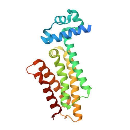
1T56

Crystal structure of TetR family repressor M. tuberculosis EthR


Experimental Data Snapshot

  • Method: X-RAY DIFFRACTION
  • Resolution: 1.70 Å
  • R-Value Free: 0.222 
  • R-Value Work: 0.208 
  • R-Value Observed: 0.208 

wwPDB Validation   3D Report Full Report


This is version 1.3 of the entry. See complete history


Literature

Crystal Structure of the TetR/CamR Family Repressor Mycobacterium tuberculosis EthR Implicated in Ethionamide Resistance.

Dover, L.G.Corsino, P.E.Daniels, I.R.Cocklin, S.L.Tatituri, V.Besra, G.S.Futterer, K.

(2004) J Mol Biol 340: 1095-1105

  • DOI: https://doi.org/10.1016/j.jmb.2004.06.003
  • Primary Citation of Related Structures:  
    1T56

  • PubMed Abstract: 

    Ethionamide has been used for more than 30 years as a second-line chemotherapeutic to treat tuberculosis patients who have developed resistance to first-line drugs, such as isoniazid (INH) and rifampicin. Activation of the pro-drug ethionamide is regulated by the Baeyer-Villiger monooxygenase EthA and the TetR/CamR family repressor EthR, whose open reading frames are separated by 75 bp on the Mycobacterium tuberculosis genome. EthR has been shown to repress transcription of the activator gene ethA by binding to this intergenic region, thus contributing to ethionamide resistance. We have determined the crystal structure of EthR, to 1.7A resolution, revealing a dimeric two-domain molecule with an overall architecture typical for TetR/CamR repressor proteins. A 20A long hydrophobic tunnel-like cavity in the "drug-binding" domain of EthR is occupied by two 1,4-dioxane molecules, a component of the crystallisation buffer. Comparing the present structure to those of the homologues Staphylococcus aureus QacR and Escherichia coli TetR leads to the hypothesis that the hydrophobic cavity constitutes a binding site for an as yet unknown ligand that might regulate DNA-binding of EthR.


  • Organizational Affiliation

    School of Biosciences, The University of Birmingham, Edgbaston, Birmingham B15 2TT, UK.


Macromolecules
Find similar proteins by:  (by identity cutoff)  |  3D Structure
Entity ID: 1
MoleculeChains Sequence LengthOrganismDetailsImage
EthR repressor216Mycobacterium tuberculosis H37RvMutation(s): 0 
UniProt
Find proteins for P9WMC1 (Mycobacterium tuberculosis (strain ATCC 25618 / H37Rv))
Explore P9WMC1 
Go to UniProtKB:  P9WMC1
Entity Groups  
Sequence Clusters30% Identity50% Identity70% Identity90% Identity95% Identity100% Identity
UniProt GroupP9WMC1
Sequence Annotations
Expand
  • Reference Sequence
Experimental Data & Validation

Experimental Data

  • Method: X-RAY DIFFRACTION
  • Resolution: 1.70 Å
  • R-Value Free: 0.222 
  • R-Value Work: 0.208 
  • R-Value Observed: 0.208 
  • Space Group: P 41 21 2
Unit Cell:
Length ( Å )Angle ( ˚ )
a = 121.5α = 90
b = 121.5β = 90
c = 33.5γ = 90
Software Package:
Software NamePurpose
REFMACrefinement
MOSFLMdata reduction
CCP4data scaling
SOLVEphasing

Structure Validation

View Full Validation Report


Entry History 

Deposition Data

Revision History  (Full details and data files)

  • Version 1.0: 2004-07-27
    Type: Initial release
  • Version 1.1: 2008-04-30
    Changes: Version format compliance
  • Version 1.2: 2011-07-13
    Changes: Advisory, Derived calculations, Source and taxonomy, Version format compliance
  • Version 1.3: 2024-02-14
    Changes: Data collection, Database references, Derived calculations