1T3L

Structural Analysis of the Voltage-Dependent Calcium Channel Beta Subunit Functional Core in Complex with Alpha1 Interaction Domain


Experimental Data Snapshot

  • Method: X-RAY DIFFRACTION
  • Resolution: 2.20 Å
  • R-Value Free: 0.282 
  • R-Value Work: 0.221 
  • R-Value Observed: 0.226 

wwPDB Validation   3D Report Full Report


This is version 1.4 of the entry. See complete history


Literature

Structural Analysis of Voltage-Dependent Calcium Channel Beta Subunit Functional Core and Its Complex with the Alpha1 Interaction Domain

Opatowsky, Y.Chen, C.-C.Campbell, K.P.Hirsch, J.A.

(2004) Neuron 42: 387-399

  • DOI: https://doi.org/10.1016/s0896-6273(04)00250-8
  • Primary Citation of Related Structures:  
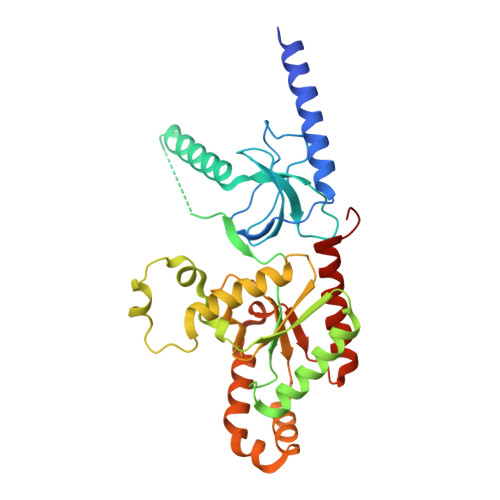
    1T3L, 1T3S

  • PubMed Abstract: 

    Voltage-dependent calcium channels (VDCC) are multiprotein assemblies that regulate the entry of extracellular calcium into electrically excitable cells and serve as signal transduction centers. The alpha1 subunit forms the membrane pore while the intracellular beta subunit is responsible for trafficking of the channel to the plasma membrane and modulation of its electrophysiological properties. Crystallographic analyses of a beta subunit functional core alone and in complex with a alpha1 interaction domain (AID) peptide, the primary binding site of beta to the alpha1 subunit, reveal that beta represents a novel member of the MAGUK protein family. The findings illustrate how the guanylate kinase fold has been fashioned into a protein-protein interaction module by alteration of one of its substrate sites. Combined results indicate that the AID peptide undergoes a helical transition in binding to beta. We outline the mechanistic implications for understanding the beta subunit's broad regulatory role of the VDCC, particularly via the AID.


  • Organizational Affiliation

    Department of Biochemistry, Faculty of Life Sciences, Tel Aviv University, Ramat Aviv 69978, Israel.


Macromolecules
Find similar proteins by:  (by identity cutoff)  |  3D Structure
Entity ID: 1
MoleculeChains Sequence LengthOrganismDetailsImage
Dihydropyridine-sensitive L-type, calcium channel beta-2 subunit337Oryctolagus cuniculusMutation(s): 1 
Gene Names: CACNB2CACNLB2
UniProt
Find proteins for P54288 (Oryctolagus cuniculus)
Explore P54288 
Go to UniProtKB:  P54288
Entity Groups  
Sequence Clusters30% Identity50% Identity70% Identity90% Identity95% Identity100% Identity
UniProt GroupP54288
Sequence Annotations
Expand
  • Reference Sequence

Find similar proteins by:  Sequence   |   3D Structure  

Entity ID: 2
MoleculeChains Sequence LengthOrganismDetailsImage
Voltage-dependent L-type calcium channel alpha-1S subunit18N/AMutation(s): 0 
UniProt
Find proteins for P07293 (Oryctolagus cuniculus)
Explore P07293 
Go to UniProtKB:  P07293
Entity Groups  
Sequence Clusters30% Identity50% Identity70% Identity90% Identity95% Identity100% Identity
UniProt GroupP07293
Sequence Annotations
Expand
  • Reference Sequence
Experimental Data & Validation

Experimental Data

  • Method: X-RAY DIFFRACTION
  • Resolution: 2.20 Å
  • R-Value Free: 0.282 
  • R-Value Work: 0.221 
  • R-Value Observed: 0.226 
  • Space Group: P 21 21 2
Unit Cell:
Length ( Å )Angle ( ˚ )
a = 72.775α = 90
b = 168.262β = 90
c = 34.233γ = 90
Software Package:
Software NamePurpose
REFMACrefinement
DENZOdata reduction
SCALEPACKdata scaling
CNSphasing

Structure Validation

View Full Validation Report


Entry History 

Deposition Data

Revision History  (Full details and data files)

  • Version 1.0: 2004-05-25
    Type: Initial release
  • Version 1.1: 2008-04-30
    Changes: Version format compliance
  • Version 1.2: 2011-07-13
    Changes: Version format compliance
  • Version 1.3: 2021-10-27
    Changes: Database references
  • Version 1.4: 2024-02-14
    Changes: Data collection