1RK2

E. COLI RIBOKINASE COMPLEXED WITH RIBOSE AND ADP, SOLVED IN SPACE GROUP P212121


Experimental Data Snapshot

  • Method: X-RAY DIFFRACTION
  • Resolution: 2.25 Å
  • R-Value Free: 0.265 
  • R-Value Work: 0.228 

wwPDB Validation   3D Report Full Report


Ligand Structure Quality Assessment 


This is version 2.1 of the entry. See complete history


Literature

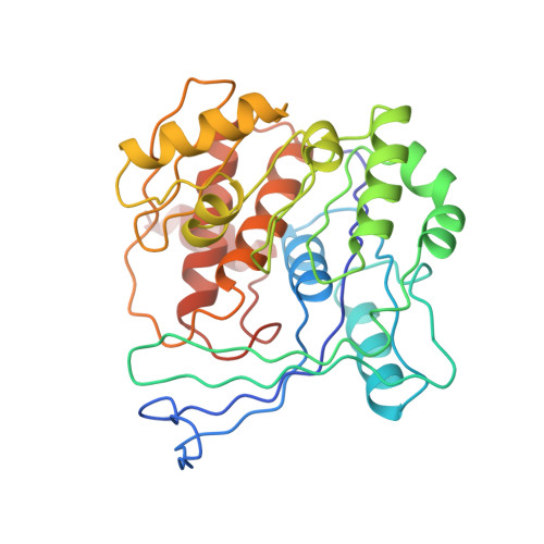
Induced fit on sugar binding activates ribokinase.

Sigrell, J.A.Cameron, A.D.Mowbray, S.L.

(1999) J Mol Biol 290: 1009-1018

  • DOI: https://doi.org/10.1006/jmbi.1999.2938
  • Primary Citation of Related Structures:  
    1RK2, 1RKA, 1RKS

  • PubMed Abstract: 

    The enzyme ribokinase phosphorylates ribose at O5* as the first step in its metabolism. The original X-ray structure of Escherichia coli ribokinase represented the ternary complex including ribose and ADP. Structures are presented here for the apo enzyme, as well as the ribose-bound state and four new ternary complex forms. Combined, the structures suggest that large and small conformational changes play critical roles in the function of this kinase. An initially open apo form can allow entry of the ribose substrate. After ribose binding, the active site lid is observed in a closed conformation, with the sugar trapped underneath. This closure and associated changes in the protein appear to assist ribokinase in recognition of the co-substrate ATP as the next step. Binding of the nucleotide brings about further, less dramatic adjustments in the enzyme structure. Additional small movements are almost certainly required during the phosphoryltransfer reaction. Evidence is presented that some types of movements of the lid are allowed in the ternary complex, which may be critical to the creation and breakdown of the transition state. Similar events are likely to take place during catalysis by other related carbohydrate kinases, including adenosine kinase.


  • Organizational Affiliation

    Department of Molecular Biology, Uppsala University, Uppsala, Sweden.


Macromolecules
Find similar proteins by:  (by identity cutoff)  |  3D Structure
Entity ID: 1
MoleculeChains Sequence LengthOrganismDetailsImage
RIBOKINASE
A, B, C, D
309Escherichia coliMutation(s): 0 
Gene Names: RBSK
EC: 2.7.1.15
UniProt
Find proteins for P0A9J6 (Escherichia coli (strain K12))
Explore P0A9J6 
Go to UniProtKB:  P0A9J6
Entity Groups  
Sequence Clusters30% Identity50% Identity70% Identity90% Identity95% Identity100% Identity
UniProt GroupP0A9J6
Sequence Annotations
Expand
  • Reference Sequence
Small Molecules
Ligands 4 Unique
IDChains Name / Formula / InChI Key2D Diagram3D Interactions
ADP
Query on ADP

Download Ideal Coordinates CCD File 
H [auth A],
L [auth B],
P [auth C],
T [auth D]
ADENOSINE-5'-DIPHOSPHATE
C10 H15 N5 O10 P2
XTWYTFMLZFPYCI-KQYNXXCUSA-N
RIB
Query on RIB

Download Ideal Coordinates CCD File 
E [auth A],
I [auth B],
M [auth C],
Q [auth D]
alpha-D-ribofuranose
C5 H10 O5
HMFHBZSHGGEWLO-AIHAYLRMSA-N
ALF
Query on ALF

Download Ideal Coordinates CCD File 
F [auth A],
J [auth B],
N [auth C],
R [auth D]
TETRAFLUOROALUMINATE ION
Al F4
UYOMQIYKOOHAMK-UHFFFAOYSA-J
MG
Query on MG

Download Ideal Coordinates CCD File 
G [auth A],
K [auth B],
O [auth C],
S [auth D]
MAGNESIUM ION
Mg
JLVVSXFLKOJNIY-UHFFFAOYSA-N
Experimental Data & Validation

Experimental Data

  • Method: X-RAY DIFFRACTION
  • Resolution: 2.25 Å
  • R-Value Free: 0.265 
  • R-Value Work: 0.228 
  • Space Group: P 21 21 21
Unit Cell:
Length ( Å )Angle ( ˚ )
a = 55.201α = 90
b = 62.492β = 90
c = 337.8γ = 90
Software Package:
Software NamePurpose
AMoREphasing
CNSrefinement
DENZOdata reduction
SCALEPACKdata scaling

Structure Validation

View Full Validation Report


Ligand Structure Quality Assessment 


Entry History 

Deposition Data

Revision History  (Full details and data files)

  • Version 1.0: 1999-09-01
    Type: Initial release
  • Version 1.1: 2008-04-26
    Changes: Version format compliance
  • Version 1.2: 2011-07-13
    Changes: Version format compliance
  • Version 2.0: 2020-07-29
    Type: Remediation
    Reason: Carbohydrate remediation
    Changes: Advisory, Atomic model, Data collection, Derived calculations, Structure summary
  • Version 2.1: 2023-08-23
    Changes: Advisory, Data collection, Database references, Refinement description, Structure summary