1RJM

Crystal Structure of MenB (Rv0548c) from Mycobacterium tuberculosis


Experimental Data Snapshot

  • Method: X-RAY DIFFRACTION
  • Resolution: 2.15 Å
  • R-Value Free: 0.231 
  • R-Value Work: 0.203 

wwPDB Validation   3D Report Full Report


Ligand Structure Quality Assessment 


This is version 1.4 of the entry. See complete history


Literature

Structure of naphthoate synthase (MenB) from Mycobacterium tuberculosis in both native and product-bound forms.

Johnston, J.M.Arcus, V.L.Baker, E.N.

(2005) Acta Crystallogr D Biol Crystallogr 61: 1199-1206

  • DOI: https://doi.org/10.1107/S0907444905017531
  • Primary Citation of Related Structures:  
    1RJM, 1RJN

  • PubMed Abstract: 

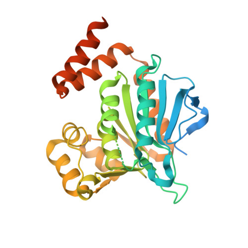
    Mycobacterium tuberculosis, the cause of tuberculosis, is one of the most devastating human pathogens. New drugs for its control are urgently needed. Menaquinone, also known as vitamin K, is an essential cofactor that is required for electron transfer and the enzymes that synthesize it are therefore potential drug targets. The enzyme naphthoate synthase (MenB) from M. tuberculosis has been expressed in Escherichia coli, purified and crystallized both as the native enzyme and in complex with naphthoyl-CoA. Both structures have been determined by X-ray crystallography: native MenB at 2.15 A resolution (R = 0.203, R(free) = 0.231) and its napthoyl-CoA complex at 2.30 A resolution (R = 0.197, R(free) = 0.225). The protein structure, which has a fold characteristic of the crotonase family of enzymes, is notable for the presence of several highly flexible regions around the active site. The bound naphthoyl-CoA is only visible for one of the three molecules in the asymmetric unit and only partly rigidifies the structure. The C-terminal region of the protein is seen to play a critical role both in completion of the binding pocket and in stabilization of the hexamer, suggesting a link between oligomerization and catalytic activity.


  • Organizational Affiliation

    School of Biological Sciences, University of Auckland, Private Bag 92019, Auckland, New Zealand.


Macromolecules
Find similar proteins by:  (by identity cutoff)  |  3D Structure
Entity ID: 1
MoleculeChains Sequence LengthOrganismDetailsImage
MenB
A, B, C
339Mycobacterium tuberculosisMutation(s): 0 
Gene Names: MenB
EC: 4.1.3.36
UniProt
Find proteins for P9WNP5 (Mycobacterium tuberculosis (strain ATCC 25618 / H37Rv))
Explore P9WNP5 
Go to UniProtKB:  P9WNP5
Entity Groups  
Sequence Clusters30% Identity50% Identity70% Identity90% Identity95% Identity100% Identity
UniProt GroupP9WNP5
Sequence Annotations
Expand
  • Reference Sequence
Small Molecules
Ligands 1 Unique
IDChains Name / Formula / InChI Key2D Diagram3D Interactions
EP1
Query on EP1

Download Ideal Coordinates CCD File 
D [auth A]3-[4-(2-HYDROXYETHYL)PIPERAZIN-1-YL]PROPANE-1-SULFONIC ACID
C9 H20 N2 O4 S
OWXMKDGYPWMGEB-UHFFFAOYSA-N
Experimental Data & Validation

Experimental Data

  • Method: X-RAY DIFFRACTION
  • Resolution: 2.15 Å
  • R-Value Free: 0.231 
  • R-Value Work: 0.203 
  • Space Group: P 43 21 2
Unit Cell:
Length ( Å )Angle ( ˚ )
a = 80.305α = 90
b = 80.305β = 90
c = 257.553γ = 90
Software Package:
Software NamePurpose
CNSrefinement
DENZOdata reduction
SCALEPACKdata scaling
AMoREphasing

Structure Validation

View Full Validation Report


Ligand Structure Quality Assessment 


Entry History 

Deposition Data

Revision History  (Full details and data files)

  • Version 1.0: 2004-11-30
    Type: Initial release
  • Version 1.1: 2008-04-29
    Changes: Version format compliance
  • Version 1.2: 2011-07-13
    Changes: Derived calculations, Version format compliance
  • Version 1.3: 2021-07-21
    Changes: Advisory, Database references, Derived calculations
  • Version 1.4: 2023-08-23
    Changes: Advisory, Data collection, Database references, Refinement description