1REA

STRUCTURE OF THE RECA PROTEIN-ADP COMPLEX


Experimental Data Snapshot

  • Method: X-RAY DIFFRACTION
  • Resolution: 2.70 Å
  • R-Value Work: 0.223 
  • R-Value Observed: 0.223 

wwPDB Validation   3D Report Full Report


This is version 1.4 of the entry. See complete history


Literature

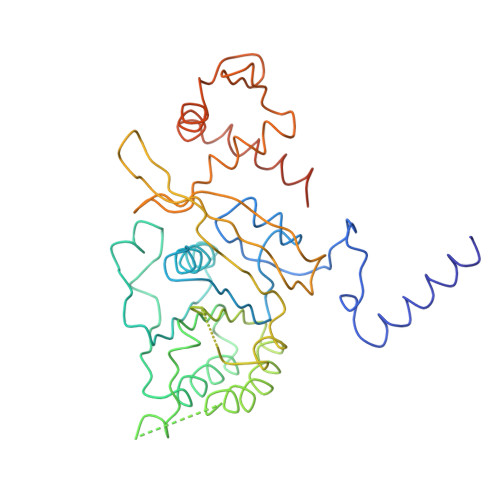
Structure of the recA protein-ADP complex.

Story, R.M.Steitz, T.A.

(1992) Nature 355: 374-376

  • DOI: https://doi.org/10.1038/355374a0
  • Primary Citation of Related Structures:  
    1REA

  • PubMed Abstract: 

    The recA protein catalyses the ATP-driven homologous pairing and strand exchange of DNA molecules. It is an allosteric enzyme: the ATPase activity is DNA-dependent, and ATP-bound recA protein has a high affinity for DNA, whereas the ADP-bound form has a low affinity. In the absence of ATP hydrolysis, recA protein can still promote homologous pairing, apparently through the formation of a triple-stranded intermediate. The exact role of ATP hydrolysis is not clear, but it presumably drives the triplex intermediate towards products. Here we determine the position of bound ADP diffused into the recA crystal. We show that only the phosphates are bound in the same way as in other NTPases containing the G/AXXXXGKT/S motif. We propose that recA protein may change its conformation upon ATP hydrolysis in a manner analogous to one such protein, the p21 protein from the ras oncogene. A model is presented to account for the allosteric stimulation of DNA binding by ATP. The mechanism by which nucleoside triphosphate hydrolysis is coupled to the binding of another ligand in recA protein and p21 may be typical of the large class of NTPases containing this conserved motif.


  • Organizational Affiliation

    Department of Molecular Biophysics and Biochemistry, Yale University, New Haven, Connecticut 06511.


Macromolecules
Find similar proteins by:  (by identity cutoff)  |  3D Structure
Entity ID: 1
MoleculeChains Sequence LengthOrganismDetailsImage
REC A352Escherichia coliMutation(s): 0 
UniProt
Find proteins for P0A7G6 (Escherichia coli (strain K12))
Explore P0A7G6 
Go to UniProtKB:  P0A7G6
Entity Groups  
Sequence Clusters30% Identity50% Identity70% Identity90% Identity95% Identity100% Identity
UniProt GroupP0A7G6
Sequence Annotations
Expand
  • Reference Sequence
Small Molecules
Ligands 1 Unique
IDChains Name / Formula / InChI Key2D Diagram3D Interactions
ADP
Query on ADP

Download Ideal Coordinates CCD File 
B [auth A]ADENOSINE-5'-DIPHOSPHATE
C10 H15 N5 O10 P2
XTWYTFMLZFPYCI-KQYNXXCUSA-N
Experimental Data & Validation

Experimental Data

  • Method: X-RAY DIFFRACTION
  • Resolution: 2.70 Å
  • R-Value Work: 0.223 
  • R-Value Observed: 0.223 
  • Space Group: P 61
Unit Cell:
Length ( Å )Angle ( ˚ )
a = 102.2α = 90
b = 102.2β = 90
c = 83.1γ = 120
Software Package:
Software NamePurpose
X-PLORmodel building
X-PLORrefinement
X-PLORphasing

Structure Validation

View Full Validation Report


Entry History 

Deposition Data

Revision History  (Full details and data files)

  • Version 1.0: 1993-10-31
    Type: Initial release
  • Version 1.1: 2008-03-24
    Changes: Version format compliance
  • Version 1.2: 2011-07-13
    Changes: Version format compliance
  • Version 1.3: 2012-05-09
    Changes: Other
  • Version 1.4: 2024-02-14
    Changes: Data collection, Database references, Derived calculations, Other