1QY9

Crystal structure of E. coli Se-MET protein YDDE


Experimental Data Snapshot

  • Method: X-RAY DIFFRACTION
  • Resolution: 2.05 Å
  • R-Value Free: 0.207 
  • R-Value Work: 0.171 
  • R-Value Observed: 0.172 

wwPDB Validation   3D Report Full Report


This is version 1.2 of the entry. See complete history


Literature

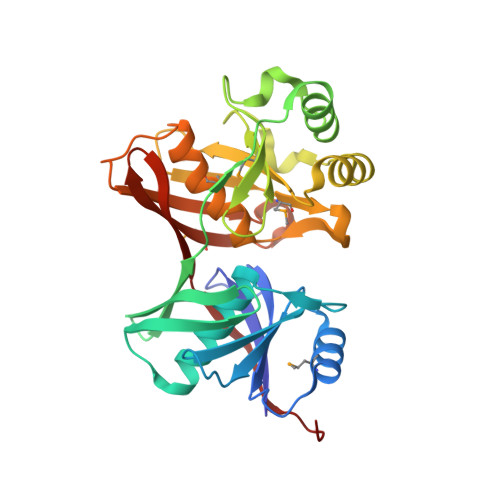
Crystal structure of E. coli yddE protein reveals a striking homology with diaminopimelate epimerase

Grassick, A.Sulzenbacher, G.Roig-Zamboni, V.Campanacci, V.Cambillau, C.Bourne, Y.

(2004) Proteins 55: 764-767


Macromolecules
Find similar proteins by:  (by identity cutoff)  |  3D Structure
Entity ID: 1
MoleculeChains Sequence LengthOrganismDetailsImage
HYPOTHETICAL PROTEIN yddE
A, B, C, D
297Escherichia coliMutation(s): 6 
Gene Names: YDDE
UniProt
Find proteins for P37757 (Escherichia coli (strain K12))
Explore P37757 
Go to UniProtKB:  P37757
Entity Groups  
Sequence Clusters30% Identity50% Identity70% Identity90% Identity95% Identity100% Identity
UniProt GroupP37757
Sequence Annotations
Expand
  • Reference Sequence
Experimental Data & Validation

Experimental Data

  • Method: X-RAY DIFFRACTION
  • Resolution: 2.05 Å
  • R-Value Free: 0.207 
  • R-Value Work: 0.171 
  • R-Value Observed: 0.172 
  • Space Group: P 1 21 1
Unit Cell:
Length ( Å )Angle ( ˚ )
a = 80.035α = 90
b = 56.529β = 101.67
c = 148.742γ = 90
Software Package:
Software NamePurpose
DENZOdata reduction
SCALAdata scaling
SOLVEphasing
RESOLVEmodel building
REFMACrefinement
CCP4data scaling
RESOLVEphasing

Structure Validation

View Full Validation Report


Entry History 

Deposition Data

Revision History  (Full details and data files)

  • Version 1.0: 2004-06-08
    Type: Initial release
  • Version 1.1: 2008-04-29
    Changes: Version format compliance
  • Version 1.2: 2011-07-13
    Changes: Advisory, Version format compliance