1QTR

CRYSTAL STRUCTURE ANALYSIS OF THE PROLYL AMINOPEPTIDASE FROM SERRATIA MARCESCENS


Experimental Data Snapshot

  • Method: X-RAY DIFFRACTION
  • Resolution: 2.32 Å
  • R-Value Free: 0.267 
  • R-Value Work: 0.189 
  • R-Value Observed: 0.189 

wwPDB Validation   3D Report Full Report


This is version 1.4 of the entry. See complete history


Literature

Crystal structure of prolyl aminopeptidase from Serratia marcescens.

Yoshimoto, T.Kabashima, T.Uchikawa, K.Inoue, T.Tanaka, N.Nakamura, K.T.Tsuru, M.Ito, K.

(1999) J Biochem 126: 559-565

  • DOI: https://doi.org/10.1093/oxfordjournals.jbchem.a022486
  • Primary Citation of Related Structures:  
    1QTR

  • PubMed Abstract: 

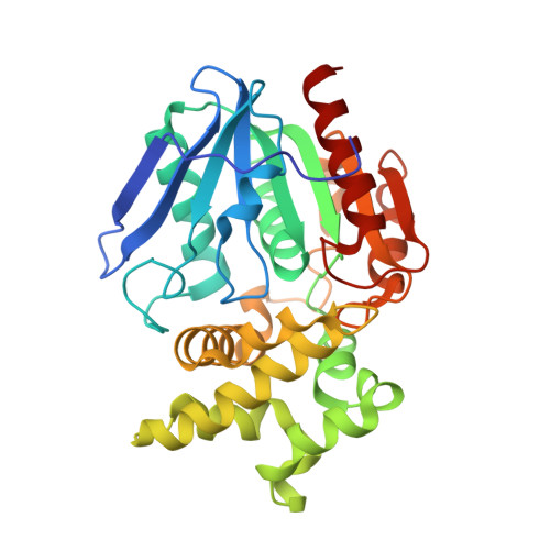
    Prolyl aminopeptidase from Serratia marcescens specifically catalyzes the removal of N-terminal proline residues from peptides. We have solved its three-dimensional structure at 2.3 A resolution by the multiple isomorphous replacement method. The enzyme consists of two contiguous domains. The larger domain shows the general topology of the alpha/beta hydrolase fold, with a central eight-stranded beta-sheet and six helices. The smaller domain consists of six helices. The catalytic triad (Ser113, His296, and Asp268) is located near the large cavity at the interface between the two domains. Cys271, which is sensitive to SH reagents, is located near the catalytic residues, in spite of the fact that the enzyme is a serine peptidase. The specific residues which make up the hydrophobic pocket line the smaller domain, and the specificity of the exo-type enzyme originates from this smaller domain, which blocks the N-terminal of P1 proline.


  • Organizational Affiliation

    School of Pharmaceutical Sciences, Nagasaki University, Nagasaki, 852-8521, Japan. t-yoshimoto@cc.nagasaki-u.ac.jp


Macromolecules
Find similar proteins by:  (by identity cutoff)  |  3D Structure
Entity ID: 1
MoleculeChains Sequence LengthOrganismDetailsImage
PROLYL AMINOPEPTIDASE317Serratia marcescensMutation(s): 0 
EC: 3.4.11.5
UniProt
Find proteins for O32449 (Serratia marcescens)
Explore O32449 
Go to UniProtKB:  O32449
Entity Groups  
Sequence Clusters30% Identity50% Identity70% Identity90% Identity95% Identity100% Identity
UniProt GroupO32449
Sequence Annotations
Expand
  • Reference Sequence
Experimental Data & Validation

Experimental Data

  • Method: X-RAY DIFFRACTION
  • Resolution: 2.32 Å
  • R-Value Free: 0.267 
  • R-Value Work: 0.189 
  • R-Value Observed: 0.189 
  • Space Group: P 43 21 2
Unit Cell:
Length ( Å )Angle ( ˚ )
a = 65.36α = 90
b = 65.36β = 90
c = 169.2γ = 90
Software Package:
Software NamePurpose
DMmodel building
X-PLORrefinement
DMphasing

Structure Validation

View Full Validation Report


Entry History 

Deposition Data

Revision History  (Full details and data files)

  • Version 1.0: 1999-07-07
    Type: Initial release
  • Version 1.1: 2008-04-27
    Changes: Version format compliance
  • Version 1.2: 2011-07-13
    Changes: Version format compliance
  • Version 1.3: 2017-10-04
    Changes: Refinement description
  • Version 1.4: 2024-02-14
    Changes: Data collection, Database references