Structure and mechanism of Sda: an inhibitor of the histidine kinases that regulate initiation of sporulation in Bacillus subtilis
Rowland, S.L., Burkholder, W.F., Cunningham, K.A., Maciejewski, M.W., Grossman, A.D., King, G.F.(2004) Mol Cell 13: 689-701
- PubMed: 15023339
- DOI: https://doi.org/10.1016/s1097-2765(04)00084-x
- Primary Citation of Related Structures:
1PV0 - PubMed Abstract:
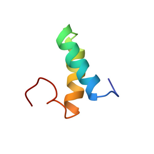
Histidine kinases are used extensively in prokaryotes to monitor and respond to changes in cellular and environmental conditions. In Bacillus subtilis, sporulation-specific gene expression is controlled by a histidine kinase phosphorelay that culminates in phosphorylation of the Spo0A transcription factor. Sda provides a developmental checkpoint by inhibiting this phosphorelay in response to DNA damage and replication defects. We show that Sda acts at the first step in the relay by inhibiting autophosphorylation of the histidine kinase KinA. The structure of Sda, which we determined using NMR, comprises a helical hairpin. A cluster of conserved residues on one face of the hairpin mediates an interaction between Sda and the KinA dimerization/phosphotransfer domain. This interaction stabilizes the KinA dimer, and the two proteins form a stable heterotetramer. The data indicate that Sda forms a molecular barricade that inhibits productive interaction between the catalytic and phosphotransfer domains of KinA.
Organizational Affiliation:
Department of Molecular, Microbial and Structural Biology, University of Connecticut Health Center, 263 Farmington Avenue, Farmington, CT 06030 USA.