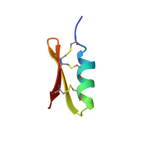
Solution structure of P05-NH2, a scorpion toxin analog with high affinity for the apamin-sensitive potassium channel.
Meunier, S., Bernassau, J.M., Sabatier, J.M., Martin-Eauclaire, M.F., Van Rietschoten, J., Cambillau, C., Darbon, H.(1993) Biochemistry 32: 11969-11976
- PubMed: 8218272
- DOI: https://doi.org/10.1021/bi00096a005
- Primary Citation of Related Structures:
1PNH - PubMed Abstract:
The venom of the scorpion Androctonus mauretanicus mauretanicus contains a toxin--P05--which is structurally and functionally similar to scorpion leiurotoxin I (87% sequence identity), a blocker of the apamin-sensitive Ca(2+)-activated K+ channels. P05, a 31-residue polypeptide cross-linked by three disulfide bridges, also possesses binding and physiological properties similar to those of the bee venom toxin apamin (18 residues, two disulfides). However, the amino acid sequences of these two polypeptides are dissimilar, except for a common Arg-Arg-Cys-Gln motif which is located on an alpha-helix. P05-NH2, a synthetic analog of P05, unlike native P05, was found to bind irreversibly to the apamin receptor. The solution structure of P05-NH2 has been solved by conventional two-dimensional NMR techniques followed by distance geometry and energy minimization. The obtained conformation is composed of two and an half turns of alpha-helix (residues 5-14) connected by a tight turn to a two-stranded antiparallel beta-sheet (sequences 17-22 and 25-29). This beta-sheet has a right-handed twist as usual for such secondary structures. The beta-turn connecting the two strands belongs to type II'. This structure is homologous to all scorpion toxin structures known so far as well as to insect defensins. The three arginines known to be involved in the pharmacological activity, i.e., Arg6, Arg7, and Arg13, are all located on the solvent-exposed side of the helix and form a positively charged surface which includes Gln9. The calculated electrostatic potential is highly asymmetric with the greatest positive potential centered on the Arg-rich alpha-helix side.(ABSTRACT TRUNCATED AT 250 WORDS)
Organizational Affiliation:
Faculté de Médecine-Nord, LCCMB, CNRS URA 1296, Marseille, France.