1P8X

The Calcium-Activated C-terminal half of gelsolin


Experimental Data Snapshot

  • Method: X-RAY DIFFRACTION
  • Resolution: 2.00 Å
  • R-Value Free: 0.245 
  • R-Value Work: 0.205 
  • R-Value Observed: 0.205 

wwPDB Validation   3D Report Full Report


This is version 1.3 of the entry. See complete history


Literature

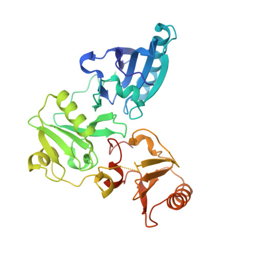
Activation in isolation: Exposure of the actin-binding site in the C-terminal half of gelsolin does not require actin

Narayan, K.Chumnarnsilpa, S.Choe, H.Irobi, E.Urosev, D.Lindberg, U.Schutt, C.E.Burtnick, L.D.Robinson, R.C.

(2003) FEBS Lett 552: 82-85

  • DOI: https://doi.org/10.1016/s0014-5793(03)00933-5
  • Primary Citation of Related Structures:  
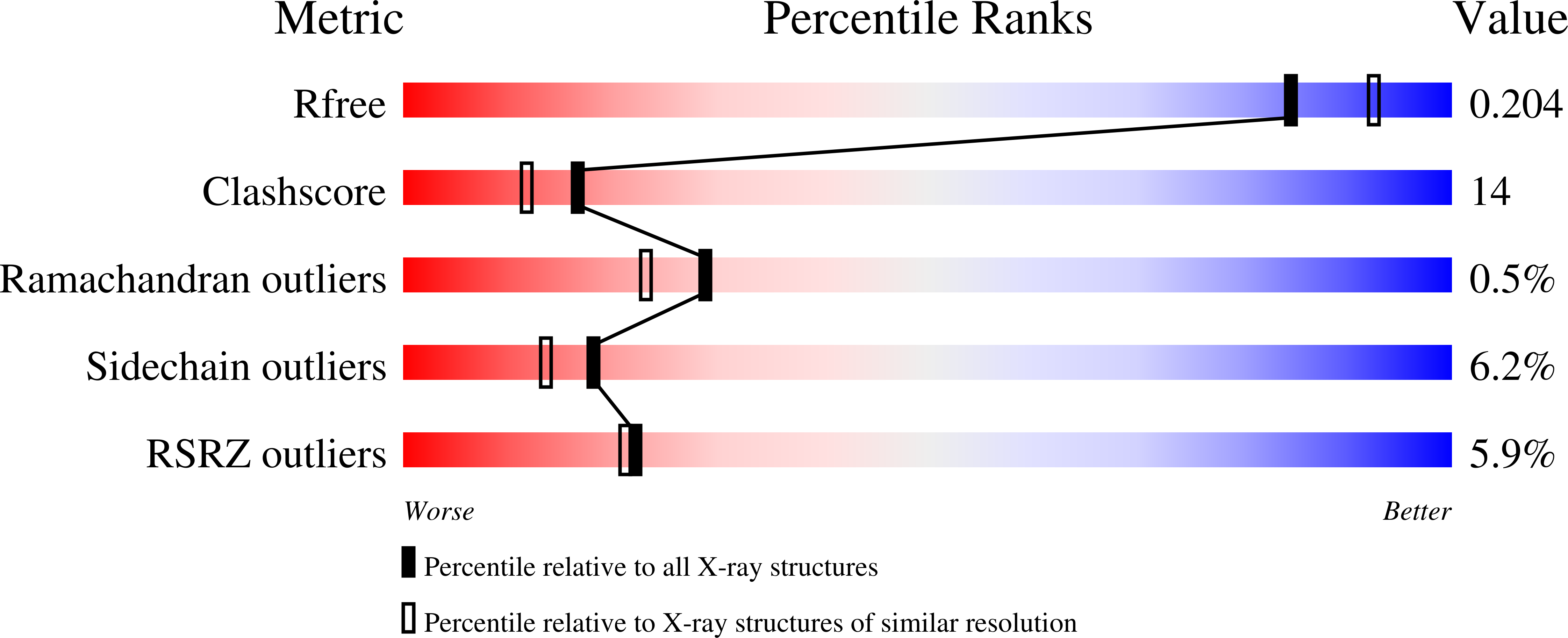
    1P8X

  • PubMed Abstract: 

    Gelsolin requires activation to carry out its severing and capping activities on F-actin. Here, we present the structure of the isolated C-terminal half of gelsolin (G4-G6) at 2.0 A resolution in the presence of Ca(2+) ions. This structure completes a triptych of the states of activation of G4-G6 that illuminates its role in the function of gelsolin. Activated G4-G6 displays an open conformation, with the actin-binding site on G4 fully exposed and all three type-2 Ca(2+) sites occupied. Neither actin nor the type-l Ca(2+), which normally is sandwiched between actin and G4, is required to achieve this conformation.


  • Organizational Affiliation

    Department of Medical Biochemistry and Microbiology, Uppsala University, Box 582, 751 23 Uppsala, Sweden.


Macromolecules
Find similar proteins by:  (by identity cutoff)  |  3D Structure
Entity ID: 1
MoleculeChains Sequence LengthOrganismDetailsImage
Gelsolin precursor, plasma
A, B, C
344Homo sapiensMutation(s): 0 
UniProt & NIH Common Fund Data Resources
Find proteins for P06396 (Homo sapiens)
Explore P06396 
Go to UniProtKB:  P06396
PHAROS:  P06396
GTEx:  ENSG00000148180 
Entity Groups  
Sequence Clusters30% Identity50% Identity70% Identity90% Identity95% Identity100% Identity
UniProt GroupP06396
Sequence Annotations
Expand
  • Reference Sequence
Experimental Data & Validation

Experimental Data

  • Method: X-RAY DIFFRACTION
  • Resolution: 2.00 Å
  • R-Value Free: 0.245 
  • R-Value Work: 0.205 
  • R-Value Observed: 0.205 
  • Space Group: P 21 21 21
Unit Cell:
Length ( Å )Angle ( ˚ )
a = 84.75α = 90
b = 90.11β = 90
c = 156.94γ = 90
Software Package:
Software NamePurpose
MOSFLMdata reduction
SCALAdata scaling
AMoREphasing
CNSrefinement
CCP4data scaling

Structure Validation

View Full Validation Report


Entry History 

Deposition Data

Revision History  (Full details and data files)

  • Version 1.0: 2003-10-14
    Type: Initial release
  • Version 1.1: 2008-04-29
    Changes: Version format compliance
  • Version 1.2: 2011-07-13
    Changes: Version format compliance
  • Version 1.3: 2023-08-16
    Changes: Data collection, Database references, Derived calculations, Refinement description