1OY0

The crystal Structure of the First Enzyme of Pantothenate Biosynthetic Pathway, Ketopantoate Hydroxymethyltransferase from Mycobacterium Tuberculosis Shows a Decameric Assembly and Terminal Helix-Swapping


Experimental Data Snapshot

  • Method: X-RAY DIFFRACTION
  • Resolution: 2.80 Å
  • R-Value Free: 0.275 
  • R-Value Work: 0.239 

wwPDB Validation   3D Report Full Report


This is version 1.3 of the entry. See complete history


Literature

The Crystal Structure of the First Enzyme in the Pantothenate Biosynthetic Pathway, Ketopantoate Hydroxymethyltransferase, from M. tuberculosis

Chaudhuri, B.N.Sawaya, M.R.Kim, C.Y.Waldo, G.S.Park, M.S.Terwilliger, T.C.Yeates, T.O.

(2003) Structure 11: 753-764

  • DOI: https://doi.org/10.1016/s0969-2126(03)00106-0
  • Primary Citation of Related Structures:  
    1OY0

  • PubMed Abstract: 

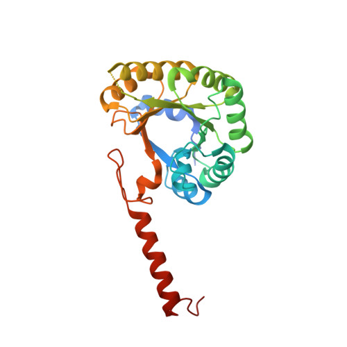
    Ketopantoate hydroxymethyltransferase (KPHMT) catalyzes the first committed step in the biosynthesis of pantothenate, which is a precursor to coenzyme A and is required for penicillin biosynthesis. The crystal structure of KPHMT from Mycobacterium tuberculosis was determined by the single anomalous substitution (SAS) method at 2.8 A resolution. KPHMT adopts a structure that is a variation on the (beta/alpha) barrel fold, with a metal binding site proximal to the presumed catalytic site. The protein forms a decameric complex, with subunits in opposing pentameric rings held together by a swapping of their C-terminal alpha helices. The structure reveals KPHMT's membership in a small, recently discovered group of (beta/alpha) barrel enzymes that employ domain swapping to form a variety of oligomeric assemblies. The apparent conservation of certain detailed structural characteristics suggests that KPHMT is distantly related by divergent evolution to enzymes in unrelated pathways, including isocitrate lyase and phosphoenolpyruvate mutase.


  • Organizational Affiliation

    UCLA Department of Chemistry and Biochemistry, University of California, Los Angeles, Los Angeles, CA 90095, USA.


Macromolecules
Find similar proteins by:  (by identity cutoff)  |  3D Structure
Entity ID: 1
MoleculeChains Sequence LengthOrganismDetailsImage
Ketopantoate hydroxymethyltransferase
A, B, C, D, E
281Mycobacterium tuberculosisMutation(s): 0 
Gene Names: PANB
EC: 2.1.2.11
UniProt
Find proteins for P9WIL7 (Mycobacterium tuberculosis (strain ATCC 25618 / H37Rv))
Explore P9WIL7 
Go to UniProtKB:  P9WIL7
Entity Groups  
Sequence Clusters30% Identity50% Identity70% Identity90% Identity95% Identity100% Identity
UniProt GroupP9WIL7
Sequence Annotations
Expand
  • Reference Sequence
Experimental Data & Validation

Experimental Data

  • Method: X-RAY DIFFRACTION
  • Resolution: 2.80 Å
  • R-Value Free: 0.275 
  • R-Value Work: 0.239 
  • Space Group: P 32 2 1
Unit Cell:
Length ( Å )Angle ( ˚ )
a = 106.8α = 90
b = 106.8β = 90
c = 224.4γ = 120
Software Package:
Software NamePurpose
DENZOdata reduction
SCALEPACKdata scaling
SOLVEphasing
DMmodel building
CNSrefinement
DMphasing

Structure Validation

View Full Validation Report


Entry History 

Revision History  (Full details and data files)

  • Version 1.0: 2003-07-15
    Type: Initial release
  • Version 1.1: 2008-04-29
    Changes: Version format compliance
  • Version 1.2: 2011-07-13
    Changes: Derived calculations, Version format compliance
  • Version 1.3: 2024-02-14
    Changes: Data collection, Database references, Derived calculations