1OP0

Crystal Structure of AaV-SP-I, a Glycosylated Snake Venom Serine Proteinase from Agkistrodon acutus


Experimental Data Snapshot

  • Method: X-RAY DIFFRACTION
  • Resolution: 2.00 Å
  • R-Value Free: 0.208 
  • R-Value Work: 0.181 
  • R-Value Observed: 0.181 

wwPDB Validation   3D Report Full Report


This is version 2.1 of the entry. See complete history


Literature

Crystal Structures and Amidolytic Activities of Two Glycosylated Snake Venom Serine Proteinases

Zhu, Z.Liang, Z.Zhang, T.Zhu, Z.Xu, W.Teng, M.Niu, L.

(2005) J Biol Chem 280: 10524-10529

  • DOI: https://doi.org/10.1074/jbc.M412900200
  • Primary Citation of Related Structures:  
    1OP0, 1OP2

  • PubMed Abstract: 

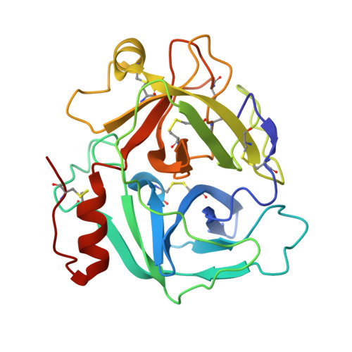
    We deduced that Agkistrodon actus venom serine proteinases I and II, previously isolated from the venom of A. acutus (Zhu, Z., Gong, P., Teng, M., and Niu, L. (2003) Acta Crystallogr. Sect. D Biol. Crystallogr. 59, 547-550), are encoded by two almost identical genes, with only the single substitution Asp for Asn at residue 62. Amidolytic assays indicated that they possess slightly different enzymatic properties. Crystal structures of A. actus venom serine proteinases I and II were determined at resolution of 2.0 and 2.1 A with the identification of trisaccharide (NAG(301)-FUC(302)-NAG(303)) and monosaccharide (NAG(301)) residues in them, respectively. The substrate binding sites S3 of the two proteinases appear much shallower than that of Trimeresurus stejnegeri venom plasminogen activator despite the overall structural similarity. Based on structural analysis, we showed that these Asn(35)-linked oligosaccharides collide spatially with some inhibitors, such as soybean trypsin inhibitor, and would therefore hinder their inhibitory binding. Difference of the carbohydrates in both the proteinases might also lead to their altered catalytic activities.


  • Organizational Affiliation

    Hefei National Laboratory for Physical Sciences at Microscale and Key Laboratory of Structural Biology, Chinese Academy of Sciences, University of Science and Technology of China, 96 Jinzhai Road, Hefei, Anhui, 230026, The People's Republic of China.


Macromolecules
Find similar proteins by:  (by identity cutoff)  |  3D Structure
Entity ID: 1
MoleculeChains Sequence LengthOrganismDetailsImage
Venom serine proteinase234Deinagkistrodon acutusMutation(s): 0 
EC: 3.4.21
UniProt
Find proteins for Q9I8X1 (Deinagkistrodon acutus)
Explore Q9I8X1 
Go to UniProtKB:  Q9I8X1
Entity Groups  
Sequence Clusters30% Identity50% Identity70% Identity90% Identity95% Identity100% Identity
UniProt GroupQ9I8X1
Sequence Annotations
Expand
  • Reference Sequence
Oligosaccharides

Help

Entity ID: 2
MoleculeChains Length2D Diagram Glycosylation3D Interactions
2-acetamido-2-deoxy-beta-D-glucopyranose-(1-4)-[alpha-L-fucopyranose-(1-6)]2-acetamido-2-deoxy-beta-D-glucopyranose
B
3N-Glycosylation
Glycosylation Resources
GlyTouCan:  G21290RB
GlyCosmos:  G21290RB
GlyGen:  G21290RB
Experimental Data & Validation

Experimental Data

  • Method: X-RAY DIFFRACTION
  • Resolution: 2.00 Å
  • R-Value Free: 0.208 
  • R-Value Work: 0.181 
  • R-Value Observed: 0.181 
  • Space Group: P 21 21 21
Unit Cell:
Length ( Å )Angle ( ˚ )
a = 49.415α = 90
b = 74.593β = 90
c = 82.419γ = 90
Software Package:
Software NamePurpose
CNSrefinement
DENZOdata reduction
SCALEPACKdata scaling
AMoREphasing

Structure Validation

View Full Validation Report


Entry History 

Deposition Data

Revision History  (Full details and data files)

  • Version 1.0: 2004-05-25
    Type: Initial release
  • Version 1.1: 2008-04-29
    Changes: Version format compliance
  • Version 1.2: 2011-07-13
    Changes: Non-polymer description, Version format compliance
  • Version 2.0: 2020-07-29
    Type: Remediation
    Reason: Carbohydrate remediation
    Changes: Atomic model, Data collection, Database references, Derived calculations, Structure summary
  • Version 2.1: 2023-10-25
    Changes: Data collection, Database references, Refinement description, Structure summary