1OMJ

CRYSTAL STRUCTURE OF A PSYCHROPHILIC ALKALINE PROTEASE FROM PSEUDOMONAS TAC II 18


Experimental Data Snapshot

  • Method: X-RAY DIFFRACTION
  • Resolution: 2.38 Å
  • R-Value Free: 0.246 
  • R-Value Work: 0.195 
  • R-Value Observed: 0.207 

wwPDB Validation   3D Report Full Report


This is version 1.6 of the entry. See complete history


Literature

Probing the role of divalent metal ions in a bacterial psychrophilic metalloprotease: binding studies of an enzyme in the crystalline state by x-ray crystallography.

Ravaud, S.Gouet, P.Haser, R.Aghajari, N.

(2003) J Bacteriol 185: 4195-4203

  • DOI: https://doi.org/10.1128/JB.185.14.4195-4203.2003
  • Primary Citation of Related Structures:  
    1O0Q, 1O0T, 1OM6, 1OM7, 1OM8, 1OMJ

  • PubMed Abstract: 

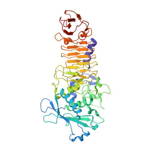
    The psychrophilic alkaline metalloprotease (PAP) produced by a Pseudomonas bacterium isolated in Antarctica belongs to the clan of metzincins, for which a zinc ion is essential for catalytic activity. Binding studies in the crystalline state have been performed by X-ray crystallography in order to improve the understanding of the role of the zinc and calcium ions bound to this protease. Cocrystallization and soaking experiments with EDTA in a concentration range from 1 to 85 mM have resulted in five three-dimensional structures with a distinct number of metal ions occupying the ion-binding sites. Evolution of the structural changes observed in the vicinity of each cation-binding site has been studied as a function of the concentration of EDTA, as well as of time, in the presence of the chelator. Among others, we have found that the catalytic zinc ion was the first ion to be chelated, ahead of a weakly bound calcium ion (Ca 700) exclusive to the psychrophilic enzyme. Upon removal of the catalytic zinc ion, the side chains of the active-site residues His-173, His-179 and Tyr-209 shifted approximately 4, 1.0, and 1.6 A, respectively. Our studies confirm and also explain the sensitivity of PAP toward moderate EDTA concentrations and propose distinct roles for the calcium ions. A new crystal form of native PAP validates our previous predictions regarding the adaptation of this enzyme to cold environments as well as the proteolytic domain calcium ion being exclusive for PAP independent of crystallization conditions.


  • Organizational Affiliation

    Laboratoire de BioCristallographie, Institut de Biologie et Chimie des Protéines, UMR 5086, CNRS-Université Claude Bernard Lyon 1, 69367 Lyon Cedex 07, France.


Macromolecules
Find similar proteins by:  (by identity cutoff)  |  3D Structure
Entity ID: 1
MoleculeChains Sequence LengthOrganismDetailsImage
SERRALYSIN463Pseudomonas sp. 'TAC II 18Mutation(s): 0 
EC: 3.4.24.40
UniProt
Find proteins for O69771 (Pseudomonas sp. 'TAC II 18')
Explore O69771 
Go to UniProtKB:  O69771
Entity Groups  
Sequence Clusters30% Identity50% Identity70% Identity90% Identity95% Identity100% Identity
UniProt GroupO69771
Sequence Annotations
Expand
  • Reference Sequence
Small Molecules
Ligands 3 Unique
IDChains Name / Formula / InChI Key2D Diagram3D Interactions
SO4
Query on SO4

Download Ideal Coordinates CCD File 
K [auth A]SULFATE ION
O4 S
QAOWNCQODCNURD-UHFFFAOYSA-L
ZN
Query on ZN

Download Ideal Coordinates CCD File 
B [auth A]ZINC ION
Zn
PTFCDOFLOPIGGS-UHFFFAOYSA-N
CA
Query on CA

Download Ideal Coordinates CCD File 
C [auth A]
D [auth A]
E [auth A]
F [auth A]
G [auth A]
C [auth A],
D [auth A],
E [auth A],
F [auth A],
G [auth A],
H [auth A],
I [auth A],
J [auth A]
CALCIUM ION
Ca
BHPQYMZQTOCNFJ-UHFFFAOYSA-N
Experimental Data & Validation

Experimental Data

  • Method: X-RAY DIFFRACTION
  • Resolution: 2.38 Å
  • R-Value Free: 0.246 
  • R-Value Work: 0.195 
  • R-Value Observed: 0.207 
  • Space Group: H 3
Unit Cell:
Length ( Å )Angle ( ˚ )
a = 184.6α = 90
b = 184.6β = 90
c = 37.85γ = 120
Software Package:
Software NamePurpose
DENZOdata reduction
SCALEPACKdata scaling
CNSrefinement
XDSdata reduction
CCP4data scaling
ROTAVATAdata scaling
AMoREphasing

Structure Validation

View Full Validation Report


Entry History 

Deposition Data

Revision History  (Full details and data files)

  • Version 1.0: 2003-07-15
    Type: Initial release
  • Version 1.1: 2008-04-29
    Changes: Version format compliance
  • Version 1.2: 2011-07-13
    Changes: Derived calculations, Version format compliance
  • Version 1.3: 2017-10-11
    Changes: Refinement description
  • Version 1.4: 2018-04-04
    Changes: Data collection
  • Version 1.5: 2018-04-11
    Changes: Data collection
  • Version 1.6: 2023-08-16
    Changes: Data collection, Database references, Derived calculations, Refinement description