Global Structure and Dynamics of Human Apolipoprotein Cii in Complex with Micelles: Evidence for Increased Mobility of the Helix Involved in the Activation of Lipoprotein Lipase
Zdunek, J., Martinez, G.V., Schleucher, J., Lycksell, P.O., Yin, Y., Nilsson, S., Shen, Y., Olivecrona, G., Wijmenga, S.(2003) Biochemistry 42: 1872
- PubMed: 12590574
- DOI: https://doi.org/10.1021/bi0267184
- Primary Citation of Related Structures:
1O8T - PubMed Abstract:
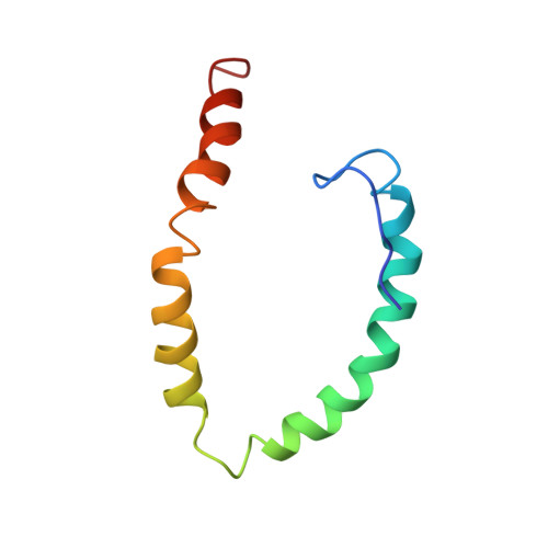
Apolipoprotein CII (apoCII), a surface constituent of plasma lipoproteins, is the activator for lipoprotein lipase (LPL) and is therefore central for lipid transport in blood. The three-dimensional structure of (13)C-, (15)N-enriched human full-length apoCII in complex with sodium dodecyl sulfate (SDS) micelles is reported. In addition to the structure determination, (15)N-relaxation measurements have been performed at two magnetic fields to characterize the dynamics of the backbone of apoCII in the complex. The relaxation data also provided global structural constraints, viz. the orientation of helices in the complex. In addition, global constraints were derived from the fact that apoCII helices are attached to the surface of the SDS micelle and that the hydrophobic moments of each helix faces the interior of the micelle. These three categories of global constraints, together with the local classical NMR constraints, were sufficient to define the 3D structure of the apoCII-SDS micelle complex. To our knowledge, this presents the first example in which the global structure of a protein-SDS micelle complex has been determined. The C-terminal helix of apoCII is known to be responsible for the activation of LPL. This helix is distinguished from the other helices by a higher degree of internal motion on the nanosecond time scale as shown by the relaxation data. The overall structure and the internal dynamics, combined with previous mutation data, give important clues toward a possible mechanism for the activation of LPL by apoCII.
Organizational Affiliation:
Department of Medical Biochemistry and Biophysics and Medical Biosciences, Physiological Chemistry, Umeå University, SE-901 87 Umeå, Sweden.