1O7F

CRYSTAL STRUCTURE OF THE REGULATORY DOMAIN OF EPAC2


Experimental Data Snapshot

  • Method: X-RAY DIFFRACTION
  • Resolution: 2.50 Å
  • R-Value Free: 0.278 
  • R-Value Work: 0.244 
  • R-Value Observed: 0.244 

wwPDB Validation   3D Report Full Report


This is version 1.2 of the entry. See complete history


Literature

Structure and Regulation of the Camp-Binding Domains of Epac2

Rehmann, H.Prakash, B.Wolf, E.Rueppel, A.De Rooij, J.Bos, J.L.Wittinghofer, A.

(2002) Nat Struct Biol 10: 26

  • DOI: https://doi.org/10.1038/nsb878
  • Primary Citation of Related Structures:  
    1O7F

  • PubMed Abstract: 

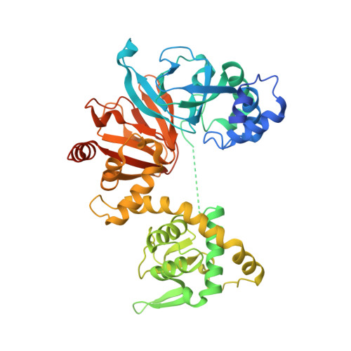
    Cyclic adenosine monophosphate (cAMP) is a universal second messenger that, in eukaryotes, was believed to act only on cAMP-dependent protein kinase A (PKA) and cyclic nucleotide-regulated ion channels. Recently, guanine nucleotide exchange factors specific for the small GTP-binding proteins Rap1 and Rap2 (Epacs) were described, which are also activated directly by cAMP. Here, we have determined the three-dimensional structure of the regulatory domain of Epac2, which consists of two cyclic nucleotide monophosphate (cNMP)-binding domains and one DEP (Dishevelled, Egl, Pleckstrin) domain. This is the first structure of a cNMP-binding domain in the absence of ligand, and comparison with previous structures, sequence alignment and biochemical experiments allow us to delineate a mechanism for cyclic nucleotide-mediated conformational change and activation that is most likely conserved for all cNMP-regulated proteins. We identify a hinge region that couples cAMP binding to a conformational change of the C-terminal regions. Mutations in the hinge of Epac can uncouple cAMP binding from its exchange activity.


  • Organizational Affiliation

    Max-Planck Institut für Molekulare Physiologie, Otto Hahn Strasse 11, D-44227, Dortmund, Germany.


Macromolecules
Find similar proteins by:  (by identity cutoff)  |  3D Structure
Entity ID: 1
MoleculeChains Sequence LengthOrganismDetailsImage
CAMP-DEPENDENT RAP1 GUANINE-NUCLEOTIDE EXCHANGE FACTOR469Mus musculusMutation(s): 0 
UniProt & NIH Common Fund Data Resources
Find proteins for Q9EQZ6 (Mus musculus)
Explore Q9EQZ6 
Go to UniProtKB:  Q9EQZ6
IMPC:  MGI:1917723
Entity Groups  
Sequence Clusters30% Identity50% Identity70% Identity90% Identity95% Identity100% Identity
UniProt GroupQ9EQZ6
Sequence Annotations
Expand
  • Reference Sequence
Experimental Data & Validation

Experimental Data

  • Method: X-RAY DIFFRACTION
  • Resolution: 2.50 Å
  • R-Value Free: 0.278 
  • R-Value Work: 0.244 
  • R-Value Observed: 0.244 
  • Space Group: P 21 21 21
Unit Cell:
Length ( Å )Angle ( ˚ )
a = 67.429α = 90
b = 96.062β = 90
c = 103.776γ = 90
Software Package:
Software NamePurpose
CNSrefinement
XDSdata reduction
XSCALEdata scaling
SOLVEphasing
SnBphasing
SHARPphasing

Structure Validation

View Full Validation Report


Entry History 

Deposition Data

Revision History  (Full details and data files)

  • Version 1.0: 2002-11-11
    Type: Initial release
  • Version 1.1: 2011-05-08
    Changes: Version format compliance
  • Version 1.2: 2011-07-13
    Changes: Version format compliance