1NWU

Crystal structure of human cartilage gp39 (HC-gp39) in complex with chitotetraose


Experimental Data Snapshot

  • Method: X-RAY DIFFRACTION
  • Resolution: 2.20 Å
  • R-Value Free: 0.245 
  • R-Value Work: 0.225 
  • R-Value Observed: 0.225 

wwPDB Validation   3D Report Full Report


This is version 2.1 of the entry. See complete history


Literature

Crystal structure and carbohydrate-binding properties of the human cartilage glycoprotein-39

Fusetti, F.Pijning, T.Kalk, K.H.Bos, E.Dijkstra, B.W.

(2003) J Biol Chem 278: 37753-37760

  • DOI: https://doi.org/10.1074/jbc.M303137200
  • Primary Citation of Related Structures:  
    1NWR, 1NWS, 1NWT, 1NWU

  • PubMed Abstract: 

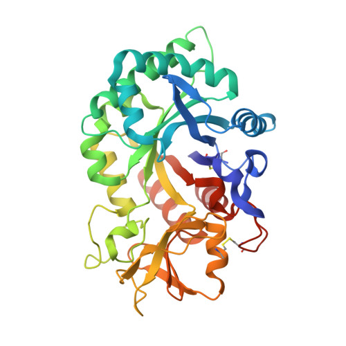
    The human cartilage glycoprotein-39 (HCgp-39 or YKL40) is expressed by synovial cells and macrophages during inflammation. Its precise physiological role is unknown. However, it has been proposed that HCgp-39 acts as an autoantigen in rheumatoid arthritis, and high expression levels have been associated with cancer development. HCgp-39 shares high sequence homology with family 18 chitinases, and although it binds to chitin it lacks enzymatic activity. The crystal structure of HCgp-39 shows that the protein displays a (beta/alpha)8-barrel fold with an insertion of an alpha + beta domain. A 43-A long carbohydrate-binding cleft is present at the C-terminal side of the beta-strands in the (beta/alpha)8 barrel. Binding of chitin fragments of different lengths identified nine sugar-binding subsites in the groove. Protein-carbohydrate interactions are mainly mediated by stacking of side chains of aromatic amino acid residues. Surprisingly, the specificity of chitin binding to HCgp-39 depends on the length of the oligosaccharide. Although chitin disaccharides tend to occupy the distal subsites, longer chains bind preferably to the central subsites in the groove. Despite the absence of enzymatic activity, long chitin fragments are distorted upon binding, with the GlcNAc at subsite -1 in a boat conformation, similar to what has been observed in chitinases. The presence of chitin in the human body has never been documented so far. However, the binding features observed in the complex structures suggest that either chitin or a closely related oligosaccharide could act as the physiological ligand for HCgp-39.


  • Organizational Affiliation

    Laboratory of Biophysical Chemistry, Department of Chemistry, University of Groningen, Nijenborgh 4, 9747AG Groningen, The Netherlands.


Macromolecules
Find similar proteins by:  (by identity cutoff)  |  3D Structure
Entity ID: 1
MoleculeChains Sequence LengthOrganismDetailsImage
Chitinase-3 like protein 1
A, B, C, D
362Homo sapiensMutation(s): 0 
UniProt & NIH Common Fund Data Resources
Find proteins for P36222 (Homo sapiens)
Explore P36222 
Go to UniProtKB:  P36222
PHAROS:  P36222
GTEx:  ENSG00000133048 
Entity Groups  
Sequence Clusters30% Identity50% Identity70% Identity90% Identity95% Identity100% Identity
UniProt GroupP36222
Sequence Annotations
Expand
  • Reference Sequence
Oligosaccharides

Help

Entity ID: 2
MoleculeChains Length2D Diagram Glycosylation3D Interactions
2-acetamido-2-deoxy-beta-D-glucopyranose-(1-4)-2-acetamido-2-deoxy-beta-D-glucopyranose
E, G, I, K
2N-Glycosylation
Glycosylation Resources
GlyTouCan:  G42666HT
GlyCosmos:  G42666HT
GlyGen:  G42666HT
Entity ID: 3
MoleculeChains Length2D Diagram Glycosylation3D Interactions
2-acetamido-2-deoxy-alpha-D-glucopyranose-(1-4)-2-acetamido-2-deoxy-beta-D-glucopyranose-(1-4)-2-acetamido-2-deoxy-beta-D-glucopyranose-(1-4)-2-acetamido-2-deoxy-beta-D-glucopyranose
F, J, L
4N/A
Glycosylation Resources
GlyTouCan:  G79211JZ
GlyCosmos:  G79211JZ
GlyGen:  G79211JZ
Entity ID: 4
MoleculeChains Length2D Diagram Glycosylation3D Interactions
2-acetamido-2-deoxy-alpha-D-glucopyranose-(1-4)-2-acetamido-2-deoxy-beta-D-glucopyranose-(1-4)-2-acetamido-2-deoxy-beta-D-glucopyranose-(1-4)-2-acetamido-2-deoxy-alpha-D-glucopyranose
H
4N/A
Glycosylation Resources
GlyTouCan:  G79253ZP
GlyCosmos:  G79253ZP
GlyGen:  G79253ZP
Experimental Data & Validation

Experimental Data

  • Method: X-RAY DIFFRACTION
  • Resolution: 2.20 Å
  • R-Value Free: 0.245 
  • R-Value Work: 0.225 
  • R-Value Observed: 0.225 
  • Space Group: P 43
Unit Cell:
Length ( Å )Angle ( ˚ )
a = 128.712α = 90
b = 128.712β = 90
c = 108.838γ = 90
Software Package:
Software NamePurpose
CNSrefinement
DENZOdata reduction
SCALEPACKdata scaling
CNSphasing

Structure Validation

View Full Validation Report


Entry History 

Deposition Data

Revision History  (Full details and data files)

  • Version 1.0: 2003-08-26
    Type: Initial release
  • Version 1.1: 2008-04-29
    Changes: Version format compliance
  • Version 1.2: 2011-07-13
    Changes: Non-polymer description, Version format compliance
  • Version 2.0: 2020-07-29
    Type: Remediation
    Reason: Carbohydrate remediation
    Changes: Atomic model, Data collection, Database references, Derived calculations, Structure summary
  • Version 2.1: 2023-11-29
    Changes: Data collection, Database references, Structure summary