1NHR

AN L40C MUTATION CONVERTS THE CYSTEINE-SULFENIC ACID REDOX CENTRE IN ENTEROCOCCAL NADH PEROXIDASE TO A DISULFIDE


Experimental Data Snapshot

  • Method: X-RAY DIFFRACTION
  • Resolution: 2.10 Å
  • R-Value Work: 0.170 
  • R-Value Observed: 0.170 

wwPDB Validation   3D Report Full Report


This is version 1.3 of the entry. See complete history


Literature

An L40C mutation converts the cysteine-sulfenic acid redox center in enterococcal NADH peroxidase to a disulfide.

Miller, H.Mande, S.S.Parsonage, D.Sarfaty, S.H.Hol, W.G.Claiborne, A.

(1995) Biochemistry 34: 5180-5190

  • DOI: https://doi.org/10.1021/bi00015a032
  • Primary Citation of Related Structures:  
    1NHR, 1NHS

  • PubMed Abstract: 

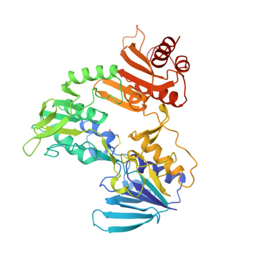
    Multiple sequence alignments including the enterococcal NADH peroxidase and NADH oxidase indicate that residues Ser38 and Cys42 align with the two cysteines of the redox-active disulfides found in glutathione reductase (GR), lipoamide dehydrogenase, mercuric reductase, and trypanothione reductase. In order to evaluate those structural determinants involved in the selection of the cysteine-sulfenic acid (Cys-SOH) redox centers found in the two peroxide reductases and the redox-active disulfides present in the GR class of disulfide reductases, NADH peroxidase residues Ser38, Phe39, Leu40, and Ser41 have been individually replaced with Cys. Both the F39C and L40C mutant peroxidases yield active-site disulfides involving the new Cys and the native Cys42; formation of the Cys39-Cys42 disulfide, however, precludes binding of the FAD coenzyme. In contrast, the L40C mutant contains tightly-bound FAD and has been analyzed by both kinetic and spectroscopic approaches. In addition, the L40C and S41C mutant structures have been determined at 2.1 and 2.0 A resolution, respectively, by X-ray crystallography. Formation of the Cys40-Cys42 disulfide bond requires a movement of Cys42-SG to a new position 5.9 A from the flavin-C(4a) position; this is consistent with the inability of the new disulfide to function as a redox center in concert with the flavin. Stereochemical constraints prohibit formation of the Cys41-Cys42 disulfide in the latter mutant.


  • Organizational Affiliation

    Department of Biochemistry, Wake Forest University Medical Center, Winston-Salem, North Carolina 27157, USA.


Macromolecules
Find similar proteins by:  (by identity cutoff)  |  3D Structure
Entity ID: 1
MoleculeChains Sequence LengthOrganismDetailsImage
NADH PEROXIDASE447Enterococcus faecalisMutation(s): 1 
EC: 1.11.1.1
UniProt
Find proteins for P37062 (Enterococcus faecalis (strain ATCC 700802 / V583))
Explore P37062 
Go to UniProtKB:  P37062
Entity Groups  
Sequence Clusters30% Identity50% Identity70% Identity90% Identity95% Identity100% Identity
UniProt GroupP37062
Sequence Annotations
Expand
  • Reference Sequence
Small Molecules
Ligands 2 Unique
IDChains Name / Formula / InChI Key2D Diagram3D Interactions
FAD
Query on FAD

Download Ideal Coordinates CCD File 
D [auth A]FLAVIN-ADENINE DINUCLEOTIDE
C27 H33 N9 O15 P2
VWWQXMAJTJZDQX-UYBVJOGSSA-N
SO4
Query on SO4

Download Ideal Coordinates CCD File 
B [auth A],
C [auth A]
SULFATE ION
O4 S
QAOWNCQODCNURD-UHFFFAOYSA-L
Experimental Data & Validation

Experimental Data

  • Method: X-RAY DIFFRACTION
  • Resolution: 2.10 Å
  • R-Value Work: 0.170 
  • R-Value Observed: 0.170 
  • Space Group: I 2 2 2
Unit Cell:
Length ( Å )Angle ( ˚ )
a = 77.7α = 90
b = 134.6β = 90
c = 145.6γ = 90
Software Package:
Software NamePurpose
X-PLORmodel building
X-PLORrefinement
X-PLORphasing

Structure Validation

View Full Validation Report


Entry History 

Deposition Data

Revision History  (Full details and data files)

  • Version 1.0: 1995-02-14
    Type: Initial release
  • Version 1.1: 2008-03-24
    Changes: Version format compliance
  • Version 1.2: 2011-07-13
    Changes: Derived calculations, Version format compliance
  • Version 1.3: 2012-03-28
    Changes: Structure summary