1NG4

Structure of ThiO (glycine oxidase) from Bacillus subtilis


Experimental Data Snapshot

  • Method: X-RAY DIFFRACTION
  • Resolution: 2.30 Å
  • R-Value Free: 0.234 
  • R-Value Work: 0.203 
  • R-Value Observed: 0.203 

wwPDB Validation   3D Report Full Report


Ligand Structure Quality Assessment 


This is version 1.4 of the entry. See complete history


Literature

Structural and Mechanistic Studies on ThiO, a Glycine Oxidase Essential for Thiamin Biosynthesis in Bacillus subtilis

Settembre, E.C.Dorrestein, P.C.Park, J.Augustine, A.Begley, T.P.Ealick, S.E.

(2003) Biochemistry 42: 2971-2981

  • DOI: https://doi.org/10.1021/bi026916v
  • Primary Citation of Related Structures:  
    1NG3, 1NG4

  • PubMed Abstract: 

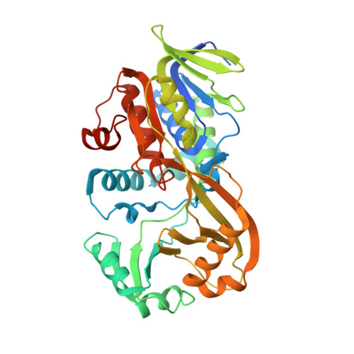
    The thiO gene of Bacillus subtilis encodes an FAD-dependent glycine oxidase. This enzyme is a homotetramer with a monomer molecular mass of 42 kDa. In this paper, we demonstrate that ThiO is required for the biosynthesis of the thiazole moiety of thiamin pyrophosphate and describe the structure of the enzyme with N-acetylglycine bound at the active site. The closest structural relatives of ThiO are sarcosine oxidase and d-amino acid oxidase. The ThiO structure, as well as the observation that N-cyclopropylglycine is a good substrate, supports a hydride transfer mechanism for the enzyme. A mechanistic proposal for the role of ThiO in thiazole biosynthesis is also described.


  • Organizational Affiliation

    Department of Chemistry and Chemical Biology, Cornell University, Ithaca, New York 14853, USA.


Macromolecules
Find similar proteins by:  (by identity cutoff)  |  3D Structure
Entity ID: 1
MoleculeChains Sequence LengthOrganismDetailsImage
Glycine oxidase
A, B
390Bacillus subtilisMutation(s): 0 
Gene Names: GOXB
EC: 1.5.3
UniProt
Find proteins for O31616 (Bacillus subtilis (strain 168))
Explore O31616 
Go to UniProtKB:  O31616
Entity Groups  
Sequence Clusters30% Identity50% Identity70% Identity90% Identity95% Identity100% Identity
UniProt GroupO31616
Sequence Annotations
Expand
  • Reference Sequence
Experimental Data & Validation

Experimental Data

  • Method: X-RAY DIFFRACTION
  • Resolution: 2.30 Å
  • R-Value Free: 0.234 
  • R-Value Work: 0.203 
  • R-Value Observed: 0.203 
  • Space Group: P 61 2 2
Unit Cell:
Length ( Å )Angle ( ˚ )
a = 139.456α = 90
b = 139.456β = 90
c = 210.278γ = 120
Software Package:
Software NamePurpose
CNSrefinement
Adxvdata processing
MOSFLMdata reduction
CCP4data scaling
CNSphasing

Structure Validation

View Full Validation Report


Ligand Structure Quality Assessment 


Entry History 

Deposition Data

Revision History  (Full details and data files)

  • Version 1.0: 2003-04-08
    Type: Initial release
  • Version 1.1: 2008-04-29
    Changes: Version format compliance
  • Version 1.2: 2011-07-13
    Changes: Derived calculations, Version format compliance
  • Version 1.3: 2017-10-11
    Changes: Refinement description
  • Version 1.4: 2024-02-14
    Changes: Data collection, Database references, Derived calculations