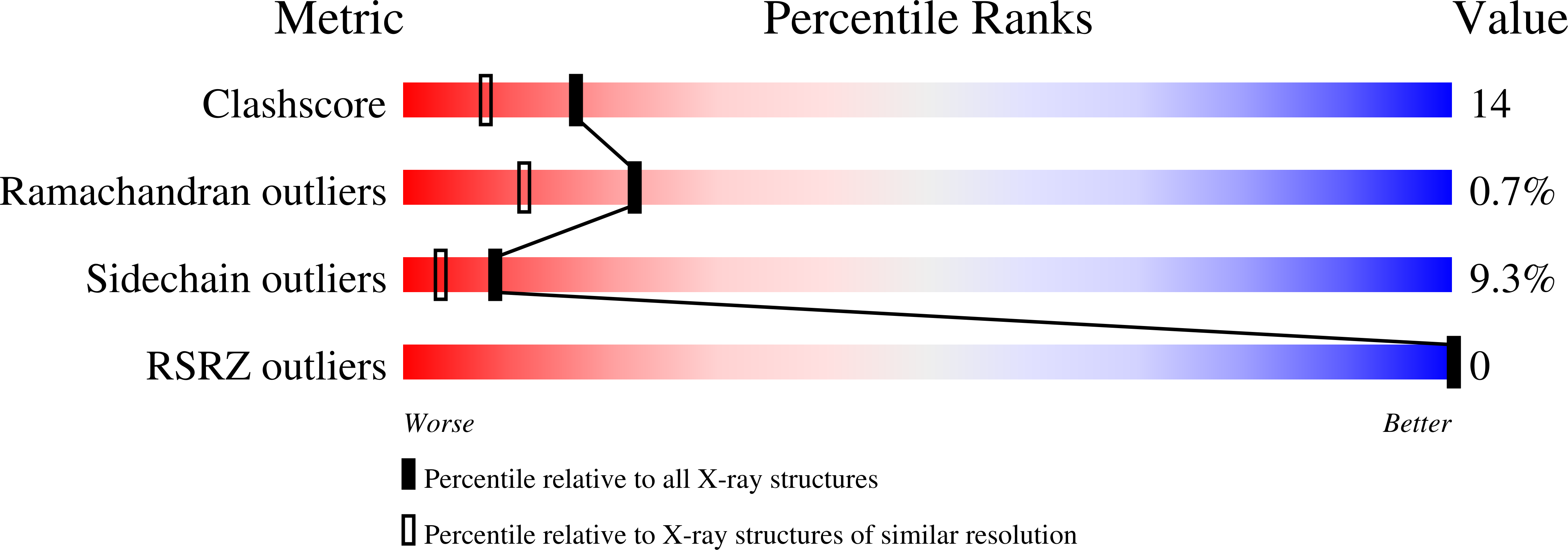
Alteration of axial coordination by protein engineering in myoglobin. Bisimidazole ligation in the His64-->Val/Val68-->His double mutant.
Dou, Y., Admiraal, S.J., Ikeda-Saito, M., Krzywda, S., Wilkinson, A.J., Li, T., Olson, J.S., Prince, R.C., Pickering, I.J., George, G.N.(1995) J Biol Chem 270: 15993-16001
- PubMed: 7608158
- DOI: https://doi.org/10.1074/jbc.270.27.15993
- Primary Citation of Related Structures:
1MNI - PubMed Abstract:
Pig and human myoglobin have been engineered to reverse the positions of the distal histidine and valine (i.e. His64(E7)-->Val and Val68(E11)-->His). Spectroscopic and ligand binding properties have been measured for human and pig H64V/V68H myoglobin, and the structure of the pig H64V/V68H double mutant has been determined to 2.07-A resolution by x-ray crystallography. The crystal structure shows that the N epsilon of His68 is located 2.3 A away from the heme iron, resulting in the formation of a hexacoordinate species. The imidazole plane of His68 is tilted relative to the heme normal; moreover it is not parallel to that of His93, in agreement with our previous proposal (Qin, J., La Mar, G. N., Dou, Y., Admiraal, S. J., and Ikeda-Saito, M. (1994) J. Biol. Chem. 269, 1083-1090). At cryogenic temperatures, the heme iron is in a low spin state, which exhibits a highly anisotropic EPR spectrum (g1 = 3.34, g2 = 2.0, and g3 < 1), quite different from that of the imidazole complex of metmyoglobin. The mean iron-nitrogen distance is 2.01 A for the low spin ferric state as determined by x-ray spectroscopy. The ferrous form of H64V/V68H myoglobin shows an optical spectrum that is similar to that of b-type cytochromes and consistent with the hexacoordinate bisimidazole hemin structure determined by the x-ray crystallography. The double mutation lowers the ferric/ferrous couple midpoint potential from +54 mV of the wild-type protein to -128 mV. Ferrous H64V/V68H myoglobin binds CO and NO to form stable complexes, but its reaction with O2 results in a rapid autooxidation to the ferric species. All of these results demonstrate that the three-dimensional positions of His64 and Val68 in the wild-type myoglobin are as important as the chemical nature of the side chains in facilitating reversible O2 binding and inhibiting autooxidation.
Organizational Affiliation:
Department of Physiology and Biophysics, Case Western Reserve University School of Medicine, Cleveland, Ohio 44106-4970, USA.


