1MHL

CRYSTAL STRUCTURE OF HUMAN MYELOPEROXIDASE ISOFORM C CRYSTALLIZED IN SPACE GROUP P2(1) AT PH 5.5 AND 20 DEG C


Experimental Data Snapshot

  • Method: X-RAY DIFFRACTION
  • Resolution: 2.25 Å
  • R-Value Work: 0.160 
  • R-Value Observed: 0.160 

wwPDB Validation   3D Report Full Report


This is version 2.0 of the entry. See complete history


Literature

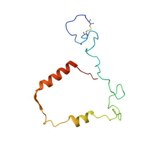
Structure of the green heme in myeloperoxidase.

Fenna, R.Zeng, J.Davey, C.

(1995) Arch Biochem Biophys 316: 653-656

  • DOI: https://doi.org/10.1006/abbi.1995.1086
  • Primary Citation of Related Structures:  
    1MHL

  • PubMed Abstract: 

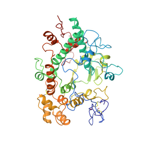
    A 3-A-resolution X-ray crystal structure of canine myeloperoxidase has previously revealed the overall structure of the molecule, including the polypeptide backbone conformation, but did not provide an unambiguous structure for the covalently bound heme. A higher resolution (2.28 A) X-ray crystal structure of human myeloperoxidase has now shown that the heme is a novel derivative of protoporphyrin IX in which three ring substituents form covalent bonds with amino acid side chains in the protein. Modified methyl groups on pyrrole rings A and C form ester linkages with glutamate 242 and aspartate 94, while a covalent bond between the vinyl group on ring A and the sulfur atom of methionine 243 results in a sulfonium ion linkage. The heme tetrapyrrole ring also shows considerable distortion from the planar conformation seen in most heme-containing proteins. The observed bending appears to result from these covalent bonds between diametrically opposed pyrrole rings A and C and the protein. Sequence comparisons suggest that the two ester linkages to the heme may also occur in other homologous mammalian peroxidases, but that the sulfonium ion linkage may be a unique feature of myeloperoxidase.


  • Organizational Affiliation

    Department of Biochemistry and Molecular Biology, University of Miami, Florida 33101.


Macromolecules
Find similar proteins by:  (by identity cutoff)  |  3D Structure
Entity ID: 1
MoleculeChains Sequence LengthOrganismDetailsImage
MYELOPEROXIDASEA,
C [auth B]
108Homo sapiensMutation(s): 0 
EC: 1.11.1.7
UniProt & NIH Common Fund Data Resources
Find proteins for P05164 (Homo sapiens)
Explore P05164 
Go to UniProtKB:  P05164
PHAROS:  P05164
GTEx:  ENSG00000005381 
Entity Groups  
Sequence Clusters30% Identity50% Identity70% Identity90% Identity95% Identity100% Identity
UniProt GroupP05164
Sequence Annotations
Expand
  • Reference Sequence
Find similar proteins by:  (by identity cutoff)  |  3D Structure
Entity ID: 2
MoleculeChains Sequence LengthOrganismDetailsImage
MYELOPEROXIDASEB [auth C],
D
466Homo sapiensMutation(s): 0 
EC: 1.11.1.7
UniProt & NIH Common Fund Data Resources
Find proteins for P05164 (Homo sapiens)
Explore P05164 
Go to UniProtKB:  P05164
PHAROS:  P05164
GTEx:  ENSG00000005381 
Entity Groups  
Sequence Clusters30% Identity50% Identity70% Identity90% Identity95% Identity100% Identity
UniProt GroupP05164
Sequence Annotations
Expand
  • Reference Sequence
Oligosaccharides

Help

Entity ID: 3
MoleculeChains Length2D Diagram Glycosylation3D Interactions
alpha-D-mannopyranose-(1-3)-[alpha-D-mannopyranose-(1-6)]beta-D-mannopyranose-(1-4)-2-acetamido-2-deoxy-beta-D-glucopyranose-(1-4)-[alpha-L-fucopyranose-(1-6)]2-acetamido-2-deoxy-beta-D-glucopyranose
E, F
6N-Glycosylation
Glycosylation Resources
GlyTouCan:  G82348BZ
GlyCosmos:  G82348BZ
GlyGen:  G82348BZ
Small Molecules
Ligands 4 Unique
IDChains Name / Formula / InChI Key2D Diagram3D Interactions
HEM
Query on HEM

Download Ideal Coordinates CCD File 
K [auth A],
P [auth B]
PROTOPORPHYRIN IX CONTAINING FE
C34 H32 Fe N4 O4
KABFMIBPWCXCRK-RGGAHWMASA-L
NAG
Query on NAG

Download Ideal Coordinates CCD File 
G [auth A],
H [auth A],
L [auth B],
M [auth B]
2-acetamido-2-deoxy-beta-D-glucopyranose
C8 H15 N O6
OVRNDRQMDRJTHS-FMDGEEDCSA-N
CA
Query on CA

Download Ideal Coordinates CCD File 
I [auth A],
N [auth B]
CALCIUM ION
Ca
BHPQYMZQTOCNFJ-UHFFFAOYSA-N
CL
Query on CL

Download Ideal Coordinates CCD File 
J [auth A],
O [auth B]
CHLORIDE ION
Cl
VEXZGXHMUGYJMC-UHFFFAOYSA-M
Experimental Data & Validation

Experimental Data

  • Method: X-RAY DIFFRACTION
  • Resolution: 2.25 Å
  • R-Value Work: 0.160 
  • R-Value Observed: 0.160 
  • Space Group: P 1 21 1
Unit Cell:
Length ( Å )Angle ( ˚ )
a = 111.7α = 90
b = 64.6β = 97.9
c = 94.2γ = 90
Software Package:
Software NamePurpose
X-PLORmodel building
X-PLORrefinement
X-PLORphasing

Structure Validation

View Full Validation Report


Entry History 

Deposition Data

Revision History  (Full details and data files)

  • Version 1.0: 1996-01-06
    Type: Initial release
  • Version 1.1: 2008-03-24
    Changes: Version format compliance
  • Version 1.2: 2011-07-13
    Changes: Derived calculations, Version format compliance
  • Version 1.3: 2017-11-29
    Changes: Advisory, Derived calculations, Other
  • Version 2.0: 2020-07-29
    Type: Remediation
    Reason: Carbohydrate remediation
    Changes: Atomic model, Data collection, Derived calculations, Structure summary