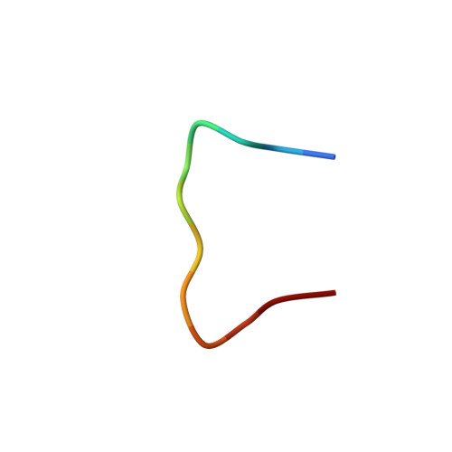
NMR structure of PW2 bound to SDS micelles. A tryptophan-rich anticoccidial peptide selected from phage display libraries
Tinoco, L.W., Da Silva Jr., A., Leite, A., Valente, A.P., Almeida, F.C.(2002) J Biol Chem 277: 36351-36356
- PubMed: 12130641
- DOI: https://doi.org/10.1074/jbc.M204225200
- Primary Citation of Related Structures:
1M02 - PubMed Abstract:
PW2 (HPLKQYWWRPSI) was selected from phage display libraries through an alternative panning method using living sporozoites of Eimeria acervulina as target. Synthetic PW2 shows anticoccidial activity against E. acervulina and Eimeria tenella with very low hemolytic activity. It also displays antifungal activity but no activity against bacteria. We present the solution structure of the PW2 bound to SDS micelles. In the absence of an interface, PW2 is in random coil conformation. In micelles, structural calculation shows that Trp-7 forms the hydrophobic core that is important for the peptide folding. Lys-4, Tyr-6, Trp-8, and Arg-9 are in the same surface, possibly facing the micelle interface. This possibility was supported by the fact that chemical shift differences for these residues were more pronounced when compared with PW2 in water and in SDS. PW2 gains structure upon binding to SDS micelles. Lys-4, Tyr-6, Trp-8, and Arg-9 were found to bind to the micelle. Trp-7, Trp-8, and Arg-9 composed the WW+ consensus found in the sequence of the peptides selected with the phage display technique against E. acervulina sporozoites. This suggested that Trp-7, Trp-8, and Arg-9 are probably key residues not only for the peptide interaction with SDS micelles but also for the interaction with E. acervulina sporozoites surface.
Organizational Affiliation:
Centro Nacional de Ressonância Magnética Nuclear, Departamento de Bioquimica Médica - Instituto de Ciencias Biomedicas, Universidade Federal do Rio de Janeiro, 21941-590 - Rio de Janeiro, RJ, Brazil.