Analysis of quinazoline and pyrido[2,3-d]pyrimidine N9-C10 reversed-bridge antifolates in complex with NADP+ and Pneumocystis carinii dihydrofolate reductase.
Cody, V., Galitsky, N., Luft, J.R., Pangborn, W., Queener, S.F., Gangjee, A.(2002) Acta Crystallogr D Biol Crystallogr 58: 1393-1399
- PubMed: 12198294
- DOI: https://doi.org/10.1107/S0907444902010442
- Primary Citation of Related Structures:
1LY3, 1LY4 - PubMed Abstract:
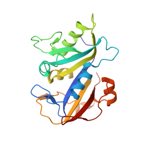
Structural studies of two ternary complexes of Pneumocystis carinii dihydrofolate reductase (pcDHFR) with the cofactor NADP(+) and potent antifolates, the N9-C10 reversed-bridge inhibitor 2,4-diamino-6-[N-(2',5'-dimethoxybenzyl)-N-methylamino]quinazoline (1) and its 3',5'-dimethoxypyrido[2,3-d]pyrimidine analog (2), were carried out. Data for the monoclinic crystals were refined to 1.90 A resolution for the complex with (1) (R = 0.178) and to 2.1 A resolution for the complex with (2) (R = 0.193). The effect of the N9-C10 reversed-bridge geometry is to distort the bridge from coplanarity with the pyrido[2,3-d]pyrimidine or quinazoline ring system and to twist the C10 methylene conformation toward a gauche conformation. This change also influences the conformation of the methoxybenzyl ring, moving it away from a trans position. This change places the 5'-methoxy group deeper within the hydrophobic pocket made by Ile65, Pro66 and Phe69 of the pcDHFR active site. These results also revealed the first observation of an unusual conformation for the reversed-bridge geometry (C5-C6-N9-C10 torsion angle) in antifolate (2). The electron density is consistent with the presence of two models (conformers 2-1 and 2-2) that result from inversion of the geometry at N9. The four examples of N9-C10 reversed-bridge antifolates cluster in two conformations, with the structure of quinazoline (1) similar to that previously reported for its 2',5'-dimethoxypyrido[2,3-d]pyrimidine analog (3). The two conformers of (2) differ from these and each other by a twisted-bridge geometry that results in the dimethoxybenzyl ring occupying the same conformational space. Conformer 2-2 also has the N9-C10 reversed bridge perpendicular to the pyrido[2,3-d]pyrimidine plane, in contrast to the gauche-trans conformation normally observed. As a result of these changes, the N9 methyl probes conformational space in the active site not normally occupied by antifolate structures. The N9 methyl of conformer 2-2 makes close contacts to the conserved Leu25 as well as the hydroxyl O atoms of the nicotinamide ribose and Ser64, whereas the other three reversed-bridge conformers make weak hydrophobic contacts with Ile123, Thr61 and Ile65. These antifolates are ten times more selective for pcDHFR than the C9-N10 bridge parent trimetrexate. However, pyrido[2,3-d]pyrimidines (2) and (3) are three times more selective for pcDHFR than quinazoline (1) is for rat liver DHFR. These data suggest that the loss of hydrogen-bonding interactions with N8 is more important to potency than the interactions of the methoxybenzyl substituents.
Organizational Affiliation:
Hauptman-Woodward Medical Research Institute, Inc., 73 High Street, Buffalo, NY 14203, USA. cody@hwi.buffalo.edu