1L8V

Crystal Structure of a Mutant (C109G,G212C) P4-P6 Domain of the Group I Intron from Tetrahymena Thermophilia


Experimental Data Snapshot

  • Method: X-RAY DIFFRACTION
  • Resolution: 2.80 Å
  • R-Value Free: 0.293 
  • R-Value Work: 0.290 
  • R-Value Observed: 0.290 

wwPDB Validation   3D Report Full Report


This is version 1.3 of the entry. See complete history


Literature

Specificity of RNA-RNA Helix Recognition

Battle, D.J.Doudna, J.A.

(2002) Proc Natl Acad Sci U S A 99: 11676-11681

  • DOI: https://doi.org/10.1073/pnas.182221799
  • Primary Citation of Related Structures:  
    1L8V

  • PubMed Abstract: 

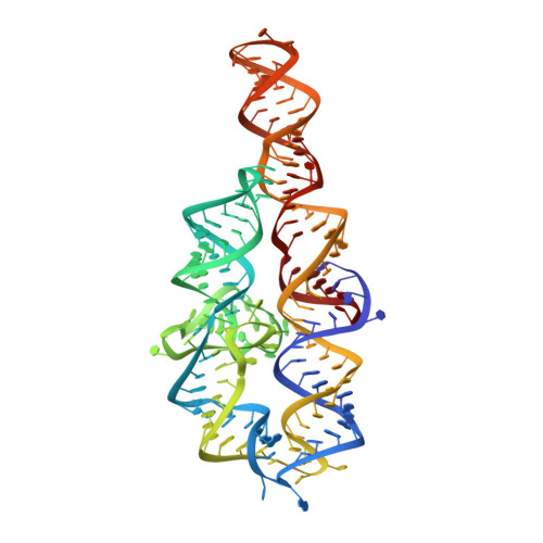
    Functional RNAs often form compact structures characterized by closely packed helices. Crystallographic analysis of several large RNAs revealed a prevalent interaction in which unpaired adenosine residues dock into the minor groove of a receptor helix. This A-minor motif, potentially the most important element responsible for global RNA architecture, has also been suggested to contribute to the fidelity of protein synthesis by discriminating against near-cognate tRNAs on the ribosome. The specificity of A-minor interactions is fundamental to RNA tertiary structure formation, as well as to their proposed role in translational accuracy. To investigate A-minor motif specificity, we analyzed mutations in an A-minor interaction within the Tetrahymena group I self-splicing intron. Thermodynamic and x-ray crystallographic results show that the A-minor interaction strongly prefers canonical base pairs over base mismatches in the receptor helix, enabling RNA interhelical packing through specific recognition of Watson-Crick minor groove geometry.


  • Organizational Affiliation

    Department of Molecular Biophysics and Biochemistry, Yale University, New Haven, CT 06520, USA.


Macromolecules
Find similar nucleic acids by:  (by identity cutoff)  |  3D Structure
Entity ID: 1
MoleculeChains LengthOrganismImage
P4-P6 RNA ribozyme domain
A, B
157N/A
Sequence Annotations
Expand
  • Reference Sequence
Experimental Data & Validation

Experimental Data

  • Method: X-RAY DIFFRACTION
  • Resolution: 2.80 Å
  • R-Value Free: 0.293 
  • R-Value Work: 0.290 
  • R-Value Observed: 0.290 
  • Space Group: P 21 21 21
Unit Cell:
Length ( Å )Angle ( ˚ )
a = 80.455α = 90
b = 129.918β = 90
c = 144.686γ = 90
Software Package:
Software NamePurpose
CNSrefinement
DENZOdata reduction
SCALEPACKdata scaling
CNSphasing

Structure Validation

View Full Validation Report


Entry History 

Deposition Data

Revision History  (Full details and data files)

  • Version 1.0: 2002-08-23
    Type: Initial release
  • Version 1.1: 2008-04-28
    Changes: Version format compliance
  • Version 1.2: 2011-07-13
    Changes: Version format compliance
  • Version 1.3: 2023-08-16
    Changes: Data collection, Database references, Derived calculations, Refinement description