1KZ8

CRYSTAL STRUCTURE OF PORCINE FRUCTOSE-1,6-BISPHOSPHATASE COMPLEXED WITH A NOVEL ALLOSTERIC-SITE INHIBITOR


Experimental Data Snapshot

  • Method: X-RAY DIFFRACTION
  • Resolution: 2.00 Å
  • R-Value Free: 0.214 
  • R-Value Work: 0.177 
  • R-Value Observed: 0.179 

wwPDB Validation   3D Report Full Report


Ligand Structure Quality Assessment 


This is version 1.4 of the entry. See complete history


Literature

ANILINOQUINAZOLINE INHIBITORS OF FRUCTOSE 1,6-BISPHOSPHATASE BIND AT A NOVEL ALLOSTERIC SITE: SYNTHESIS, IN VITRO CHARACTERIZATION, AND X-RAY CRYSTALLOGRAPHY

Wright, S.W.Carlo, A.A.Carty, M.D.Danley, D.E.Hageman, D.L.Karam, G.A.Levy, C.B.Mansour, M.N.Mathiowetz, A.M.McClure, L.D.Nestor, N.B.McPherson, R.K.Pandit, J.Pustilnik, L.R.Schulte, G.K.Soeller, W.C.Treadway, J.L.Wang, I.-K.Bauer, P.H.

(2002) J Med Chem 45: 3865-3877

  • DOI: https://doi.org/10.1021/jm010496a
  • Primary Citation of Related Structures:  
    1KZ8

  • PubMed Abstract: 

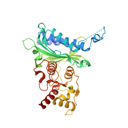
    The synthesis and in vitro structure-activity relationships (SAR) of a novel series of anilinoquinazolines as allosteric inhibitors of fructose-1,6-bisphosphatase (F16Bpase) are reported. The compounds have a different SAR as inhibitors of F16Bpase than anilinoquinazolines previously reported. Selective inhibition of F16Bpase can be attained through the addition of appropriate polar functional groups at the quinazoline 2-position, thus separating the F16Bpase inhibitory activity from the epidermal growth factor receptor tyrosine kinase inhibitory activity previously observed with similar structures. The compounds have been found to bind at a symmetry-repeated novel allosteric site at the subunit interface of the enzyme. Inhibition is brought about by binding to a loop comprised of residues 52-72, preventing the necessary participation of these residues in the assembly of the catalytic site. Mutagenesis studies have identified the key amino acid residues in the loop that are required for inhibitor recognition and binding.


  • Organizational Affiliation

    Pfizer Central Research, Eastern Point Road, Groton, Connecticut 06340, USA. stephen_w_wright@groton.pfizer.com


Macromolecules
Find similar proteins by:  (by identity cutoff)  |  3D Structure
Entity ID: 1
MoleculeChains Sequence LengthOrganismDetailsImage
FRUCTOSE-1,6-BISPHOSPHATASEA,
B [auth F]
337Sus scrofaMutation(s): 0 
EC: 3.1.3.11
UniProt
Find proteins for P00636 (Sus scrofa)
Explore P00636 
Go to UniProtKB:  P00636
Entity Groups  
Sequence Clusters30% Identity50% Identity70% Identity90% Identity95% Identity100% Identity
UniProt GroupP00636
Sequence Annotations
Expand
  • Reference Sequence
Small Molecules
Ligands 4 Unique
IDChains Name / Formula / InChI Key2D Diagram3D Interactions
PFE
Query on PFE

Download Ideal Coordinates CCD File 
F [auth A],
J [auth F]
{4-[3-(6,7-DIETHOXY-QUINAZOLIN-4-YLAMINO)-PHENYL]-THIAZOL-2-YL}-METHANOL
C22 H22 N4 O3 S
ZJESXGUODSBHSK-UHFFFAOYSA-N
AMP
Query on AMP

Download Ideal Coordinates CCD File 
E [auth A],
I [auth F]
ADENOSINE MONOPHOSPHATE
C10 H14 N5 O7 P
UDMBCSSLTHHNCD-KQYNXXCUSA-N
F6P
Query on F6P

Download Ideal Coordinates CCD File 
C [auth A],
G [auth F]
6-O-phosphono-beta-D-fructofuranose
C6 H13 O9 P
BGWGXPAPYGQALX-ARQDHWQXSA-N
MN
Query on MN

Download Ideal Coordinates CCD File 
D [auth A],
H [auth F]
MANGANESE (II) ION
Mn
WAEMQWOKJMHJLA-UHFFFAOYSA-N
Binding Affinity Annotations 
IDSourceBinding Affinity
AMP BindingDB:  1KZ8 IC50: min: 140, max: 9800 (nM) from 10 assay(s)
PFE BindingDB:  1KZ8 IC50: min: 340, max: 1.00e+4 (nM) from 3 assay(s)
Binding MOAD:  1KZ8 IC50: 830 (nM) from 1 assay(s)
PDBBind:  1KZ8 IC50: 830 (nM) from 1 assay(s)
Experimental Data & Validation

Experimental Data

  • Method: X-RAY DIFFRACTION
  • Resolution: 2.00 Å
  • R-Value Free: 0.214 
  • R-Value Work: 0.177 
  • R-Value Observed: 0.179 
  • Space Group: P 21 21 2
Unit Cell:
Length ( Å )Angle ( ˚ )
a = 59.65α = 90
b = 165.94β = 90
c = 79.223γ = 90
Software Package:
Software NamePurpose
DENZOdata reduction
SCALEPACKdata scaling
CCP4model building
REFMACrefinement
CCP4phasing

Structure Validation

View Full Validation Report


Ligand Structure Quality Assessment 


Entry History 

Revision History  (Full details and data files)

  • Version 1.0: 2002-10-16
    Type: Initial release
  • Version 1.1: 2008-04-28
    Changes: Version format compliance
  • Version 1.2: 2011-07-13
    Changes: Derived calculations, Version format compliance
  • Version 1.3: 2020-07-29
    Type: Remediation
    Reason: Carbohydrate remediation
    Changes: Data collection, Derived calculations, Structure summary
  • Version 1.4: 2023-08-16
    Changes: Data collection, Database references, Refinement description, Structure summary