1KSZ

ENTRAPMENT OF 6-THIOPHOSPHORYL-IMP IN THE ACTIVE SITE OF CRYSTALLINE ADENYLOSUCCINATE SYNTHETASE FROM ESCHERICHIA COLI, DATA COLLECTED AT 298K


Experimental Data Snapshot

  • Method: X-RAY DIFFRACTION
  • Resolution: 2.80 Å
  • R-Value Free: 0.250 
  • R-Value Work: 0.171 
  • R-Value Observed: 0.171 

wwPDB Validation   3D Report Full Report


This is version 1.4 of the entry. See complete history


Literature

Entrapment of 6-thiophosphoryl-IMP in the active site of crystalline adenylosuccinate synthetase from Escherichia coli.

Poland, B.W.Bruns, C.Fromm, H.J.Honzatko, R.B.

(1997) J Biol Chem 272: 15200-15205

  • DOI: https://doi.org/10.1074/jbc.272.24.15200
  • Primary Citation of Related Structures:  
    1KSZ, 1NHT

  • PubMed Abstract: 

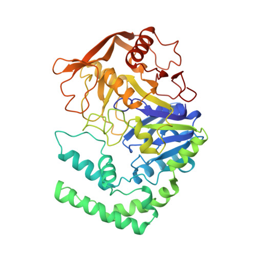
    Crystal structures of adenylosuccinate synthetase from Escherichia coli complexed with Mg2+, 6-thiophosphoryl-IMP, GDP, and hadacidin at 298 and 100 K have been refined to R-factors of 0.171 and 0.206 against data to 2.8 and 2.5 A resolution, respectively. Interactions of GDP, Mg2+ and hadacidin are similar to those observed for the same ligands in the complex of IMP, GDP, NO3-, Mg2+ and hadacidin (Poland, B. W., Fromm, H. J. & Honzatko, R. B. (1996). J. Mol. Biol. 264, 1013-1027). Although crystals were grown from solutions containing 6-mercapto-IMP and GTP, the electron density at the active site is consistent with 6-thiophosphoryl-IMP and GDP. Asp-13 and Gln-224 probably work in concert to stabilize the 6-thioanion of 6-mercapto-IMP, which in turn is the nucleophile in the displacement of GDP from the gamma-phosphate of GTP. Once formed, 6-thiophosphoryl-IMP is stable in the active site of the enzyme under the conditions of the structural investigation. The direct observation of 6-thiophosphoryl-IMP in the active site is consistent with the putative generation of 6-phosphoryl-IMP along the reaction pathway of the synthetase.


  • Organizational Affiliation

    Department of Biochemistry and Biophysics, Iowa State University, Ames, Iowa 50011, USA.


Macromolecules
Find similar proteins by:  (by identity cutoff)  |  3D Structure
Entity ID: 1
MoleculeChains Sequence LengthOrganismDetailsImage
ADENYLOSUCCINATE SYNTHETASE431Escherichia coliMutation(s): 0 
EC: 6.3.4.4
UniProt
Find proteins for P0A7D4 (Escherichia coli (strain K12))
Explore P0A7D4 
Go to UniProtKB:  P0A7D4
Entity Groups  
Sequence Clusters30% Identity50% Identity70% Identity90% Identity95% Identity100% Identity
UniProt GroupP0A7D4
Sequence Annotations
Expand
  • Reference Sequence
Small Molecules
Ligands 4 Unique
IDChains Name / Formula / InChI Key2D Diagram3D Interactions
PGS
Query on PGS

Download Ideal Coordinates CCD File 
E [auth A]2-DEAZO-6-THIOPHOSPHATE GUANOSINE-5'-MONOPHOSPHATE
C10 H15 N4 O10 P2 S
NYBPOGVUIOPXHI-KQYNXXCUSA-O
GDP
Query on GDP

Download Ideal Coordinates CCD File 
C [auth A]GUANOSINE-5'-DIPHOSPHATE
C10 H15 N5 O11 P2
QGWNDRXFNXRZMB-UUOKFMHZSA-N
HDA
Query on HDA

Download Ideal Coordinates CCD File 
D [auth A]HADACIDIN
C3 H5 N O4
URJHVPKUWOUENU-UHFFFAOYSA-N
MG
Query on MG

Download Ideal Coordinates CCD File 
B [auth A]MAGNESIUM ION
Mg
JLVVSXFLKOJNIY-UHFFFAOYSA-N
Experimental Data & Validation

Experimental Data

  • Method: X-RAY DIFFRACTION
  • Resolution: 2.80 Å
  • R-Value Free: 0.250 
  • R-Value Work: 0.171 
  • R-Value Observed: 0.171 
  • Space Group: P 32 2 1
Unit Cell:
Length ( Å )Angle ( ˚ )
a = 81.63α = 90
b = 81.63β = 90
c = 159.22γ = 120
Software Package:
Software NamePurpose
X-PLORmodel building
X-PLORrefinement
XENGENdata reduction
X-PLORphasing

Structure Validation

View Full Validation Report


Entry History 

Deposition Data

Revision History  (Full details and data files)

  • Version 1.0: 1997-10-08
    Type: Initial release
  • Version 1.1: 2008-03-24
    Changes: Version format compliance
  • Version 1.2: 2011-07-13
    Changes: Version format compliance
  • Version 1.3: 2017-11-29
    Changes: Derived calculations, Other
  • Version 1.4: 2024-02-14
    Changes: Data collection, Database references, Derived calculations