Crystal structure analysis of the exocytosis-sensitive phosphoprotein, pp63/parafusin (phosphoglucomutase), from Paramecium reveals significant conformational variability.
Muller, S., Diederichs, K., Breed, J., Kissmehl, R., Hauser, K., Plattner, H., Welte, W.(2002) J Mol Biol 315: 141-153
- PubMed: 11779235
- DOI: https://doi.org/10.1006/jmbi.2001.5168
- Primary Citation of Related Structures:
1KFI, 1KFQ - PubMed Abstract:
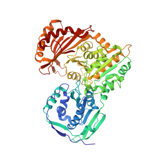
During exocytosis of dense-core secretory vesicles (trichocysts) in Paramecium, the protein pp63/parafusin (pp63/pf) is transiently dephosphorylated. We report here the structures of two crystal forms of one isoform of this protein which has a high degree of homology with rabbit phosphoglucomutase, whose structure has been reported. As expected, both proteins possess highly similar structures, showing the same four domains forming two lobes with an active-site crevice in between. The two X-ray structures that we report here were determined after crystallization in the presence of sulfate and tartrate, and show the lobes arranged as a closed and an open conformation, respectively. While both conformations possess a bound divalent cation, only the closed (sulfate-bound) conformation shows bound sulfate ions in the "phosphate-transfer site" near the catalytic serine residue and in the "phosphate-binding site". Comparison with the open form shows that the latter dianion is placed in the centre of three arginine residues, one contributed by subunit II and two by subunit IV, suggesting that it causes a contraction of the arginine triangle, which establishes the observed conformational closure of the lobes. It is therefore likely that the closed conformation forms only when a phosphoryl group is bound to the phosphate-binding site. The previously published structure of rabbit phosphoglucomutase is intermediate between these two conformers. Several of the known reversible phosphorylation sites of pp63/pf-1 are at positions critical for transition between the conformations and for binding of the ligands and thus give hints as to possible roles of pp63/pf-1 in the course of exocytosis.
Organizational Affiliation:
Department of Biology, University of Konstanz, Konstanz, 78457, Germany.