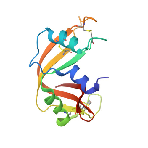
Cleavage of 3',5'-Pyrophosphate-Linked Dinucleotides by Ribonuclease A and Angiogenin
Jardine, A.M., Leonidas, D.D., Jenkins, J.L., Park, C., Raines, R.T., Acharya, K.R., Shapiro, R.(2001) Biochemistry 40: 10262-10272
- PubMed: 11513604
- DOI: https://doi.org/10.1021/bi010888j
- Primary Citation of Related Structures:
1JN4 - PubMed Abstract:
Recently, 3',5'-pyrophosphate-linked 2'-deoxyribodinucleotides were shown to be >100-fold more effective inhibitors of RNase A superfamily enzymes than were the corresponding monophosphate-linked (i.e., standard) dinucleotides. Here, we have investigated two ribo analogues of these compounds, cytidine 3'-pyrophosphate (P'-->5') adenosine (CppA) and uridine 3'-pyrophosphate (P'-->5') adenosine (UppA), as potential substrates for RNase A and angiogenin. CppA and UppA are cleaved efficiently by RNase A, yielding as products 5'-AMP and cytidine or uridine cyclic 2',3'-phosphate. The k(cat)/K(m) values are only 4-fold smaller than for the standard dinucleotides CpA and UpA, and the K(m) values (10-16 microM) are lower than those reported for any earlier small substrates (e.g., 500-700 microM for CpA and UpA). The k(cat)/K(m) value for CppA with angiogenin is also only severalfold smaller than for CpA, but the effect of lengthening the internucleotide linkage on K(m) is more modest. Ribonucleotide 3',5'-pyrophosphate linkages were proposed previously to exist in nature as chemically labile intermediates in the pathway for the generation of cyclic 2',3'-phosphate termini in various RNAs. We demonstrate that in fact they are relatively stable (t(1/2) > 15 days for uncatalyzed degradation of UppA at pH 6 and 25 degrees C) and that cleavage in vivo is most likely enzymatic. Replacements of the RNase A catalytic residues His12 and His119 by alanine reduce activity toward UppA by approximately 10(5)-and 10(3.3)-fold, respectively. Thus, both residues play important roles. His12 probably acts as a base catalyst in cleavage of UppA (as with RNA). However, the major function of His119 in RNA cleavage, protonation of the 5'-O leaving group, is not required for UppA cleavage because the pK(a) of the leaving group is much lower than that for RNA substrates. A crystal structure of the complex of RNase A with 2'-deoxyuridine 3'-pyrophosphate (P'-->5') adenosine (dUppA), determined at 1.7 A resolution, together with models of the UppA complex based on this structure suggest that His119 contributes to UppA cleavage through a hydrogen bond with a nonbridging oxygen atom in the pyrophosphate and through pi-pi stacking with the six-membered ring of adenine.
Organizational Affiliation:
Center for Biochemical and Biophysical Sciences and Medicine, Harvard Medical School, Boston, Massachusetts 02115, USA.