1JAD

C-terminal Domain of Turkey PLC-beta


Experimental Data Snapshot

  • Method: X-RAY DIFFRACTION
  • Resolution: 2.40 Å
  • R-Value Free: 0.295 
  • R-Value Work: 0.254 
  • R-Value Observed: 0.256 

wwPDB Validation   3D Report Full Report


This is version 1.2 of the entry. See complete history


Literature

A unique fold of phospholipase C-beta mediates dimerization and interaction with G alpha q.

Singer, A.U.Waldo, G.L.Harden, T.K.Sondek, J.

(2002) Nat Struct Biol 9: 32-36

  • DOI: https://doi.org/10.1038/nsb731
  • Primary Citation of Related Structures:  
    1JAD

  • PubMed Abstract: 

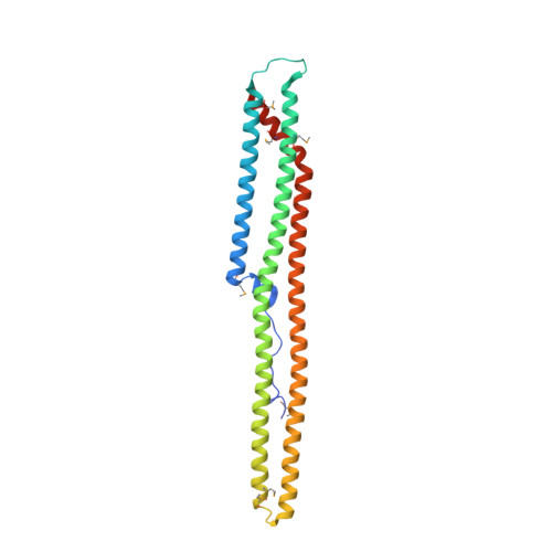
    GTP-bound subunits of the Gq family of G alpha subunits directly activate phospholipase C-beta (PLC-beta) isozymes to produce the second messengers inositol 1,4,5-trisphosphate and diacylglycerol. PLC-betas are GTPase activating proteins (GAPs) that also promote the formation of GDP-bound, inactive G beta subunits. Both phospholipase activation by G alpha-GTP subunits and GAP activity require a C-terminal region unique to PLC-beta isozymes. The crystal structure of the C-terminal region from an avian PLC-beta, determined at 2.4 A resolution, reveals a novel fold composed almost entirely of three long helices forming a coiled-coil that dimerizes along its long axis in an antiparallel orientation. The dimer interface is extensive ( approximately 3,200 A(2)), and, based on gel exclusion chromatography, full length PLC-betas are dimeric, indicating that PLC-betas likely function as dimers. Sequence conservation, mutational data and molecular modeling show that an electrostatically positive surface of the dimer contains the major determinants for binding G beta q. Effector dimerization, as highlighted by PLC-betas, provides a viable mechanism for regulating signaling cascades linked to heterotrimeric G proteins.


  • Organizational Affiliation

    Department of Pharmacology, The University of North Carolina at Chapel Hill, North Carolina 27599, USA.


Macromolecules
Find similar proteins by:  (by identity cutoff)  |  3D Structure
Entity ID: 1
MoleculeChains Sequence LengthOrganismDetailsImage
phospholipase C beta
A, B
251Meleagris gallopavoMutation(s): 7 
EC: 3.1.4.11
UniProt
Find proteins for Q91086 (Meleagris gallopavo)
Explore Q91086 
Go to UniProtKB:  Q91086
Entity Groups  
Sequence Clusters30% Identity50% Identity70% Identity90% Identity95% Identity100% Identity
UniProt GroupQ91086
Sequence Annotations
Expand
  • Reference Sequence
Small Molecules
Ligands 1 Unique
IDChains Name / Formula / InChI Key2D Diagram3D Interactions
SO4
Query on SO4

Download Ideal Coordinates CCD File 
C [auth A]SULFATE ION
O4 S
QAOWNCQODCNURD-UHFFFAOYSA-L
Modified Residues  1 Unique
IDChains TypeFormula2D DiagramParent
MSE
Query on MSE
A, B
L-PEPTIDE LINKINGC5 H11 N O2 SeMET
Experimental Data & Validation

Experimental Data

  • Method: X-RAY DIFFRACTION
  • Resolution: 2.40 Å
  • R-Value Free: 0.295 
  • R-Value Work: 0.254 
  • R-Value Observed: 0.256 
  • Space Group: P 1
Unit Cell:
Length ( Å )Angle ( ˚ )
a = 39.111α = 101.07
b = 51.928β = 96.96
c = 79.35γ = 100.7
Software Package:
Software NamePurpose
CNSrefinement
SCALEPACKdata scaling
MLPHAREphasing

Structure Validation

View Full Validation Report


Entry History 

Deposition Data

Revision History  (Full details and data files)

  • Version 1.0: 2001-12-28
    Type: Initial release
  • Version 1.1: 2008-04-27
    Changes: Version format compliance
  • Version 1.2: 2011-07-13
    Changes: Version format compliance