1IWB

Crystal structure of diol dehydratase


Experimental Data Snapshot

  • Method: X-RAY DIFFRACTION
  • Resolution: 1.85 Å
  • R-Value Free: 0.256 
  • R-Value Work: 0.181 
  • R-Value Observed: 0.181 

wwPDB Validation   3D Report Full Report


This is version 1.4 of the entry. See complete history


Literature

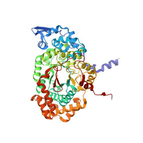
Substrate-induced conformational change of a coenzyme B12-dependent enzyme: crystal structure of the substrate-free form of diol dehydratase

Shibata, N.Masuda, J.Morimoto, Y.Yasuoka, N.Toraya, T.

(2002) Biochemistry 41: 12607-12617

  • DOI: https://doi.org/10.1021/bi026104z
  • Primary Citation of Related Structures:  
    1IWB

  • PubMed Abstract: 

    Substrate binding triggers catalytic radical formation through the cobalt-carbon bond homolysis in coenzyme B12-dependent enzymes. We have determined the crystal structure of the substrate-free form of Klebsiella oxytoca diol dehydratase*cyanocobalamin complex at 1.85 A resolution. The structure contains two units of the heterotrimer consisting of alpha, beta, and gamma subunits. As compared with the structure of its substrate-bound form, the beta subunits are tilted by approximately 3 degrees and cobalamin is also tilted so that pyrrole rings A and D are significantly lifted up toward the substrate-binding site, whereas pyrrole rings B and C are only slightly lifted up. The structure revealed that the potassium ion in the substrate-binding site of the substrate-free enzyme is also heptacoordinated; that is, two oxygen atoms of two water molecules coordinate to it instead of the substrate hydroxyls. A modeling study in which the structures of both the cobalamin moiety and the adenine ring of the coenzyme were superimposed onto those of the enzyme-bound cyanocobalamin and the adenine ring-binding pocket, respectively, demonstrated that the distortions of the Co-C bond in the substrate-free form are already marked but slightly smaller than those in the substrate-bound form. It was thus strongly suggested that the Co-C bond becomes largely activated (labilized) when the coenzyme binds to the apoenzyme even in the absence of substrate and undergoes homolysis through the substrate-induced conformational changes of the enzyme. Kinetic coupling of Co-C bond homolysis with hydrogen abstraction from the substrate shifts the equilibrium to dissociation.


  • Organizational Affiliation

    Department of Life Science, Graduate School of Science, Himeji Institute of Technology, 3-2-1 Kouto, Kamigori, Ako-gun, Hyogo 678-1297, Japan.


Macromolecules
Find similar proteins by:  (by identity cutoff)  |  3D Structure
Entity ID: 1
MoleculeChains Sequence LengthOrganismDetailsImage
DIOL DEHYDRATASE alpha chainA,
D [auth L]
554Klebsiella oxytocaMutation(s): 0 
EC: 4.2.1.28
UniProt
Find proteins for Q59470 (Klebsiella oxytoca)
Explore Q59470 
Go to UniProtKB:  Q59470
Entity Groups  
Sequence Clusters30% Identity50% Identity70% Identity90% Identity95% Identity100% Identity
UniProt GroupQ59470
Sequence Annotations
Expand
  • Reference Sequence
Find similar proteins by:  (by identity cutoff)  |  3D Structure
Entity ID: 2
MoleculeChains Sequence LengthOrganismDetailsImage
DIOL DEHYDRATASE beta chain
B, E
224Klebsiella oxytocaMutation(s): 0 
EC: 4.2.1.28
UniProt
Find proteins for Q59471 (Klebsiella oxytoca)
Explore Q59471 
Go to UniProtKB:  Q59471
Entity Groups  
Sequence Clusters30% Identity50% Identity70% Identity90% Identity95% Identity100% Identity
UniProt GroupQ59471
Sequence Annotations
Expand
  • Reference Sequence
Find similar proteins by:  (by identity cutoff)  |  3D Structure
Entity ID: 3
MoleculeChains Sequence LengthOrganismDetailsImage
DIOL DEHYDRATASE gamma chainC [auth G],
F [auth M]
173Klebsiella oxytocaMutation(s): 0 
EC: 4.2.1.28
UniProt
Find proteins for Q59472 (Klebsiella oxytoca)
Explore Q59472 
Go to UniProtKB:  Q59472
Entity Groups  
Sequence Clusters30% Identity50% Identity70% Identity90% Identity95% Identity100% Identity
UniProt GroupQ59472
Sequence Annotations
Expand
  • Reference Sequence
Experimental Data & Validation

Experimental Data

  • Method: X-RAY DIFFRACTION
  • Resolution: 1.85 Å
  • R-Value Free: 0.256 
  • R-Value Work: 0.181 
  • R-Value Observed: 0.181 
  • Space Group: P 21 21 21
Unit Cell:
Length ( Å )Angle ( ˚ )
a = 75.79α = 90
b = 122.4β = 90
c = 207.59γ = 90
Software Package:
Software NamePurpose
DENZOdata reduction
SCALEPACKdata scaling
CNSrefinement
SHELXL-97refinement
CNSphasing

Structure Validation

View Full Validation Report


Entry History 

Deposition Data

Revision History  (Full details and data files)

  • Version 1.0: 2003-05-01
    Type: Initial release
  • Version 1.1: 2008-04-27
    Changes: Version format compliance
  • Version 1.2: 2011-07-13
    Changes: Version format compliance
  • Version 1.3: 2012-10-24
    Changes: Non-polymer description
  • Version 1.4: 2023-05-31
    Changes: Database references, Derived calculations