1IRH

The Solution Structure of The Third Kunitz Domain of Tissue Factor Pathway Inhibitor


Experimental Data Snapshot

  • Method: SOLUTION NMR
  • Conformers Calculated: 40 
  • Conformers Submitted: 20 
  • Selection Criteria: all calculated structures submitted,back calculated data agree with experimental NOESY spectrum,structures with acceptable covalent geometry,structures with favorable non-bond energy,structures with the least restraint violations,structures with the lowest energy,target function 

wwPDB Validation   3D Report Full Report


This is version 1.4 of the entry. See complete history


Literature

Structural mechanism for heparin-binding of the third Kunitz domain of human tissue factor pathway inhibitor.

Mine, S.Yamazaki, T.Miyata, T.Hara, S.Kato, H.

(2002) Biochemistry 41: 78-85

  • DOI: https://doi.org/10.1021/bi011299g
  • Primary Citation of Related Structures:  
    1IRH

  • PubMed Abstract: 

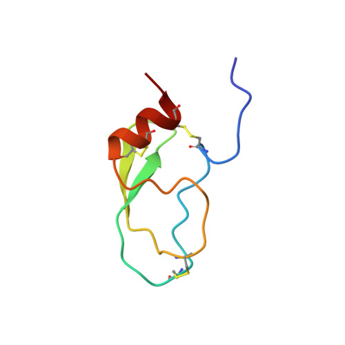
    Tissue factor pathway inhibitor (TFPI) inhibits the activity of coagulation factor VIIa and Xa through its K1 and K2 domain, respectively, and the inhibitory activity is enhanced by heparin. The function of the K3 domain of TFPI has not been established, but the domain probably harbors a heparin binding site (HBS-2). We determined the three-dimensional solution structure of the TFPI K3 domain (Glu182-Gly242) by heteronuclear multidimensional NMR. The results showed that the molecule is composed of one antiparallel beta-sheet and one alpha-helix, and in overall structure is very similar to the K2 domain, with the rms deviation of 1.55 A for the 58 defined C(alpha) positions. However, the surface electrostatic properties of both domains are different each other. The lack of inhibitory activity of the K3 domain is explained by the absence of electrostatic interaction with factor Xa over a large surface area. A titration experiment with size-fractionated heparin showed that a heparin binding site was located in the vicinity of the alpha-helix. In this region, a positively charged cluster is formed by Lys213, Lys232, and Lys240, and the negatively charged sulfate groups of heparin bind there. The enhancement of inhibitory activity by heparin probably was not due to a conformational change to TFPI itself. It is likely that heparin simply increases the local concentration of TFPI on the cell surface and stabilizes the initial complex that forms.


  • Organizational Affiliation

    National Cardiovascular Center Research Institute, 5 Fujishirodai, Suita, Osaka 565-8565 Japan.


Macromolecules
Find similar proteins by:  (by identity cutoff)  |  3D Structure
Entity ID: 1
MoleculeChains Sequence LengthOrganismDetailsImage
tissue factor pathway inhibitor61Homo sapiensMutation(s): 0 
UniProt & NIH Common Fund Data Resources
Find proteins for P10646 (Homo sapiens)
Explore P10646 
Go to UniProtKB:  P10646
PHAROS:  P10646
GTEx:  ENSG00000003436 
Entity Groups  
Sequence Clusters30% Identity50% Identity70% Identity90% Identity95% Identity100% Identity
UniProt GroupP10646
Sequence Annotations
Expand
  • Reference Sequence
Experimental Data & Validation

Experimental Data

  • Method: SOLUTION NMR
  • Conformers Calculated: 40 
  • Conformers Submitted: 20 
  • Selection Criteria: all calculated structures submitted,back calculated data agree with experimental NOESY spectrum,structures with acceptable covalent geometry,structures with favorable non-bond energy,structures with the least restraint violations,structures with the lowest energy,target function 

Structure Validation

View Full Validation Report


Entry History 

Deposition Data

Revision History  (Full details and data files)

  • Version 1.0: 2002-02-06
    Type: Initial release
  • Version 1.1: 2008-04-27
    Changes: Version format compliance
  • Version 1.2: 2011-07-13
    Changes: Version format compliance
  • Version 1.3: 2022-02-23
    Changes: Database references, Derived calculations
  • Version 1.4: 2023-12-27
    Changes: Data collection