Structure of rat transthyretin (rTTR) complex with thyroxine at 2.5 A resolution: first non-biased insight into thyroxine binding reveals different hormone orientation in two binding sites.
Wojtczak, A., Cody, V., Luft, J.R., Pangborn, W.(2001) Acta Crystallogr D Biol Crystallogr 57: 1061-1070
- PubMed: 11468389
- DOI: https://doi.org/10.1107/s0907444901007235
- Primary Citation of Related Structures:
1IE4 - PubMed Abstract:
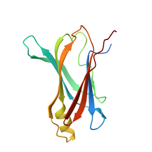
The first observation of the unique environment for thyroxine (T(4)) binding in tetrameric rat transthyretin (rTTR) is reported as determined by X-ray diffraction. These data revealed different modes of hormone binding in the two unique hormone-binding sites in the rat TTR tetramer channel. Differences in the orientation of thyroxine and the position of water molecules in the two binding sites further suggest a mechanism for the docking pathway of the hormone into the channel of TTR. Crystals of the rat transthyretin-thyroxine complex are isomorphous with those reported for apo rTTR and crystallized in the tetragonal space group P4(3)2(1)2 with four independent TTR monomeric subunits in the asymmetric part of the crystal lattice. Data were collected to 2.5 A resolution and the structure was refined to R = 20.9% for 15 384 data in the resolution range 12-2.5 A. Similar to human TTR, the rat protein is also a 54 000 Da tetramer with four identical polypeptide chains of 127 amino-acid residues. Of the 22 amino-acid residues which differ between the human and rat sequences, none are in the thyroxine-binding domains. Analysis of these structural data reveals that the tertiary structure is similar to that of hTTR, with only small differences in the flexible loop regions on the surface of the structure. Conformational changes of the amino acids in the channel result in a hydrogen-bonded network that connects the two binding domains, in contrast to the hydrogen bonds formed along the tetramer interface in the apo transthyretin structure. These changes suggest a mechanism for the signal transmission between thyroxine-binding domains.
Organizational Affiliation:
Institute of Chemistry, Nicolas Copernicus University, 87-100 Torun, Poland.